Choosing the right display technology for radiology can feel overwhelming, with technical terms like IPS and VA clouding the decision. Yet, this choice is critical, as the wrong panel could subtly distort images, potentially leading to missed diagnoses or inconsistencies in patient assessment.a company deeply invested in advancing medical display technology, I’ve seen firsthand how panel choice directly impacts clinical confidence. The good news is that understanding the core differences can clearly illuminate the superior option for diagnostic precision.
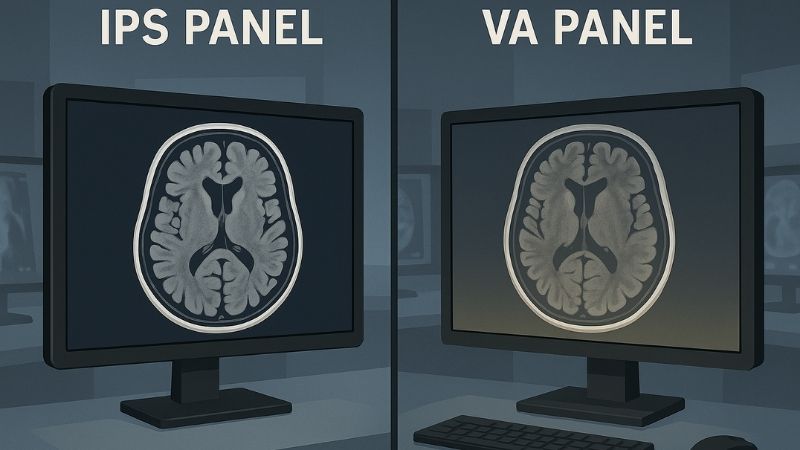
For radiology, IPS (In-Plane Switching) panels are generally better than VA (Vertical Alignment) panels. IPS technology offers superior color accuracy, wider and more consistent viewing angles, and better grayscale fidelity when viewed off-axis. These characteristics are crucial for accurate diagnostic interpretation and maintaining DICOM compliance, areas where VA panels can exhibit limitations like gamma shift and color distortion at wider angles.
This isn’t just a minor technical preference; it’s about ensuring diagnostic integrity. Let’s delve deeper into why these differences matter so much in a clinical setting.
What are the core differences between IPS and VA panels?
Navigating the technical jargon of display panels can be confusing for medical professionals. You might worry that choosing incorrectly could compromise the very images you rely on for critical diagnoses. It’s essential to demystify these terms.
The core differences lie in their liquid crystal alignment: IPS crystals align parallel to the screen, offering wide viewing angles and consistent colors, while VA crystals align perpendicularly, typically providing higher contrast but narrower optimal viewing angles.
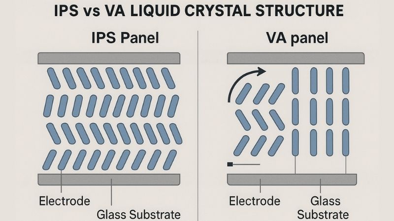
At Reshin, our mission to provide high-quality display solutions necessitates a deep understanding of these fundamental technologies. IPS (In-Plane Switching)1 and VA (Vertical Alignment)2 are two dominant types of LCD (Liquid Crystal Display) panel technologies3, and their differences stem primarily from how their liquid crystals are structured and behave when an electric current is applied.
IPS Panels:
In an IPS panel, the liquid crystals are aligned parallel to the glass substrates. When a voltage is applied, these crystals rotate in the same plane (hence "in-plane switching") to allow light from the backlight to pass through. This molecular arrangement results in:
- Superior Viewing Angles: Light is dispersed more evenly, so colors and brightness remain remarkably consistent even when viewing the screen from extreme angles (often up to 178/178 degrees).
- Excellent Color Reproduction: IPS panels are known for their accurate and consistent color fidelity across the entire screen and from various viewpoints.
VA Panels:
In a VA panel, the liquid crystals are aligned perpendicularly (vertically) to the glass substrates in their default state, effectively blocking light. When voltage is applied, they tilt to allow light through. This structure leads to:
- High Native Contrast Ratios: VA panels can achieve deeper blacks because the perpendicular crystals are very effective at blocking backlight bleed when displaying dark scenes.
- Potentially Slower Response Times: Historically, VA panels could have slightly slower pixel response times than IPS, though this gap has narrowed with modern advancements.
- Viewing Angle Limitations: While better than older TN (Twisted Nematic) panels, VA panels can suffer from "gamma shift" or "black crush" when viewed off-center – meaning dark shades can lose detail or colors can shift.
| Feature | IPS (In-Plane Switching) | VA (Vertical Alignment) |
|---|---|---|
| Crystal Alignment | Parallel to screen surface | Perpendicular to screen surface |
| Viewing Angles | Excellent, wide (up to 178°/178°) | Good, but can show shifts off-center |
| Color Accuracy | Very high and consistent | Good, but can shift at angles |
| Contrast Ratio | Good (typically 1000:1 to 1500:1) | Excellent (typically 3000:1 or higher) |
| Grayscale Fidelity | Very stable across viewing angles | Can exhibit off-axis gamma shift |
| Response Time | Generally fast | Can be slightly slower, improving |
My insight is that while VA’s high contrast seems appealing, the consistency and accuracy of IPS are far more critical for radiology.
How do viewing angles impact diagnostic accuracy in radiology?
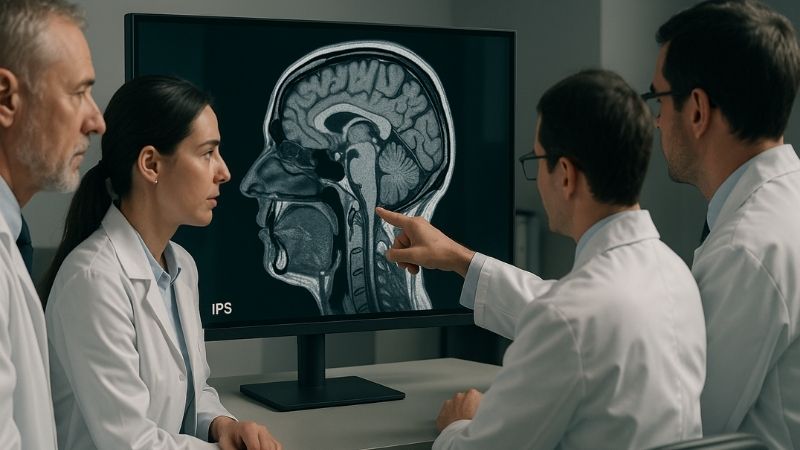
Imagine multiple specialists huddled around a monitor discussing a complex case. If the image changes depending on where each person is standing, critical details could be misinterpreted, leading to diagnostic uncertainty or error. This is a serious concern.
Poor viewing angles cause color, contrast, and brightness shifts when viewing off-center. This can lead to misinterpretation of subtle pathologies, especially when multiple clinicians consult on the same image, directly impacting diagnostic accuracy.
In a radiology department, monitors are often viewed by more than one person simultaneously, or by a single radiologist who may shift their position during a long reading session.extensive experience in operating theatre equipment integration and image processing, would understand the paramount importance of every viewer seeing the exact same image. This is where the viewing angle characteristics of IPS panels4 truly shine.
As I noted in my insights, VA panels can suffer from gamma shift5 and color distortion6 when viewed off-center. Gamma shift refers to a change in the luminance curve, meaning that the brightness relationship between different grayscale levels changes. Darker grays might appear even darker ("black crush"), obscuring subtle details in shadowed areas, or mid-tones might shift, altering the perceived texture or density of tissues. Color distortion means that hues can change or lose saturation when viewed from an angle.
Consider these scenarios where wide, consistent viewing angles are critical:
- Collaborative Diagnosis: When a senior radiologist consults with a junior colleague, or when specialists from different disciplines (e.g., oncology, surgery) review images together, they must all see the identical image presentation, regardless of their position relative to the screen. IPS panels ensure this shared visual truth.
- Teaching Environments: In academic hospitals, medical students and residents learn by observing and discussing images. Consistent image quality from all angles is essential for effective teaching.
- Radiologist Movement: Radiologists don’t always sit perfectly centered in front of the monitor. They may lean back, shift side-to-side, or stand up. An IPS display ensures the image remains stable and accurate throughout these movements. Our Reshin dual-screen diagnostic monitors like the MD45C or MD46C benefit greatly from IPS technology, as a radiologist often scans across two large displays.
The superior viewing angle performance of IPS technology directly translates to higher diagnostic confidence and reduced risk of errors stemming from display limitations.
Does panel type affect grayscale fidelity and DICOM compliance?
DICOM Part 14 is the cornerstone of consistent medical image display. Radiologists rely on this standard. If the chosen panel technology inherently struggles to maintain this standard across different viewing conditions, then diagnostic accuracy is at risk.
Yes, panel type significantly affects grayscale fidelity and DICOM compliance. IPS panels generally maintain more consistent grayscale representation across various viewing angles, crucial for adherence to the DICOM Part 14 standard, unlike VA panels which can exhibit off-axis shifts.
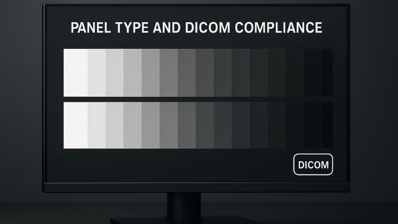
The DICOM (Digital Imaging and Communications in Medicine) Part 14 Grayscale Standard Display Function (GSDF) is designed to ensure that medical images are displayed with perceptually linearized grayscales. This means that equal changes in pixel values result in equally perceived changes in brightness, allowing for consistent interpretation of subtle tissue differences. Maintaining this standard is paramount, and panel technology plays a key role.
that IPS technology maintains consistent luminance and contrast across the entire panel. This is critical because DICOM calibration is typically performed from a central viewing position.
- IPS and DICOM: Because IPS panels exhibit minimal shift in brightness, contrast, and color when viewed from different angles, the DICOM GSDF7 calibration remains effective across a much wider field of view. This means that the subtle distinctions between different shades of gray, crucial for detecting pathologies, are preserved whether you are directly in front of the screen or slightly off to the side. This consistency is vital for models like our Reshin MD33G 3MP Grayscale Diagnostic Monitor or the high-resolution MD52G 5MP Grayscale Mammography Monitor, where detecting the faintest lesion is key.
- VA and DICOM Challenges: With VA panels8, even if perfectly DICOM calibrated from the center, the aforementioned gamma shift and contrast changes at wider angles can mean the display is no longer perceptually linearized for an off-axis viewer. A specific shade of gray might appear differently to someone viewing from the side compared to someone viewing from the center, potentially leading to inconsistent interpretations or missed details.
Furthermore, as I pointed out in my fourth insight, while VA panels boast higher native contrast ratios (deeper blacks), this advantage is often less critical in diagnostic imaging than calibrated grayscale performance and consistency. Diagnostic decisions rely on the accurate representation of many Just Noticeable Differences (JNDs) across the entire grayscale range, not just the deepest blacks. IPS technology9, with its stable characteristics, provides a more reliable platform for achieving and maintaining accurate DICOM GSDF conformance across practical viewing conditions.
Why is color consistency crucial in medical imaging?
While much of traditional radiology focuses on grayscale images, the role of color is increasingly important. If colors are not displayed accurately and consistently, then clinical information conveyed by color can be misinterpreted, potentially impacting diagnoses or treatment planning.
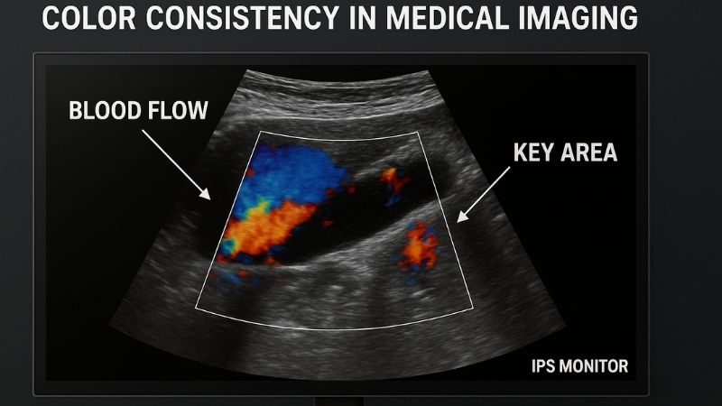
Color consistency is crucial for modalities like color Doppler ultrasound, PET-CT fusion imaging, and 3D visualizations where color encodes vital physiological or anatomical information. Inaccurate or shifting colors can lead to diagnostic errors.
I’ve seen the evolution of medical imaging towards greater use of multi-modal techniques where color plays a significant diagnostic role. While our grayscale diagnostic monitors like the MD26GA are workhorses, the demand for accurate color representation is growing.
Here’s why color consistency, a hallmark of IPS panels, is vital:
- Color Doppler Ultrasound10: In these studies, color is used to represent blood flow direction and velocity. Accurate and consistent hues (e.g., specific shades of red for flow towards the transducer, blue for flow away) are essential for assessing vascular conditions. IPS panels ensure these colors are rendered faithfully without shifting due to viewing angle. Reshin color diagnostic monitors like the MD22CA 2MP monitor or the larger MD26C 24" Diagnostic Monitor are designed for such applications.
- Fusion Imaging11: Techniques like PET-CT overlay functional (PET) images, often color-coded to show metabolic activity, onto anatomical (CT) images. The precise color values and their registration are critical. IPS displays ensure the color information from the PET scan is accurately and consistently represented.
- 3D Visualization and Surgical Planning: Many modern imaging systems create 3D reconstructions where different structures or pathologies can be color-coded for clarity. Surgeons and radiologists rely on these colors for planning interventions.
- Digital Pathology12: As pathology slides are increasingly digitized and viewed on monitors, accurate color reproduction becomes paramount for identifying cellular structures and stains.
- Multimodal Workstations: Radiologists often review images from various modalities on the same display or adjacent displays. Consistent color representation across all these images is necessary for an integrated diagnostic view. Models like our MD50C 5MP Color Mammography Monitor or the versatile 8MP Diagnostic Display benefit immensely from the color capabilities of IPS technology.
My first insight correctly states that IPS panels offer superior color consistency. This isn’t just about aesthetically pleasing images; it’s about ensuring that diagnostic information encoded in color is reliably conveyed to the clinician.
Why does Reshin prefer IPS panels in its diagnostic monitors?
Manufacturers make specific technology choices for their products. For medical displays, these choices are not arbitrary; they are driven by the stringent demands of clinical accuracy, reliability, and user confidence. Understanding this rationale helps in appreciating the product’s value.
Reshin prefers IPS panels for its diagnostic monitors because they provide the optimal combination of wide viewing angles, superior color accuracy, stable grayscale fidelity for DICOM conformance, and overall image consistency essential for confident and precise diagnoses.

Our decision at Reshin to predominantly use IPS technology13 in our diagnostic monitor14 lineup, from the entry-level MD10C 1MP Diagnostic Monitor to the advanced MD120C 12MP High-Precision Diagnostic Monitor with AI Calibration, is a deliberate one, rooted in our mission to advance medical display technology and provide high-quality solutions. My fifth insight summarizes this: "Reshin diagnostic displays adopt IPS panels due to their precise color reproduction, wide visibility range, and stability across time—ensuring radiologists can work with confidence and accuracy."
Here’s a breakdown of why IPS aligns so well with the needs of radiology professionals like Dr. Amy Chen:
- Uncompromised Viewing Experience: Radiologists need to trust that the image is accurate regardless of their viewing position or if multiple people are viewing. IPS delivers this.
- Diagnostic Precision in Grayscale and Color: Whether it’s differentiating subtle grayscale variations in a mammogram on our MD52G or interpreting color flow in an ultrasound on our MD51CHY 34" 5MP Diagnostic Monitor for X-ray Imaging, IPS provides the necessary fidelity.
- Robust DICOM Compliance15: The inherent stability of IPS panels makes it easier to achieve and maintain DICOM Part 14 conformance over the life of the monitor, which is a cornerstone of our quality promise.
- Long-Term Reliability: IPS panels have a proven track record of durability and stable performance, which is critical for the demanding environment of a hospital. Features like intelligent backlight sensors, common in our diagnostic range, work in concert with the stable IPS panel to ensure lasting image quality.
While VA panels do offer higher static contrast ratios, as mentioned in my fourth insight, this single advantage is outweighed by the potential for off-axis viewing issues that can compromise diagnostic accuracy in a typical radiology workflow. The consistent, calibrated performance of an IPS panel, even if its on-paper contrast spec is slightly lower, provides a more reliable and trustworthy image for making critical clinical decisions. This commitment to quality and reliability is why leading global brands trust Reshin and why our displays are used in over 100,000 hospitals worldwide.
Conclusion
For the demands of radiology, IPS panel technology offers superior viewing angles, color accuracy, and grayscale consistency, crucial for diagnostic confidence and DICOM compliance, making it the preferred choice over VA panels. To explore IPS-based diagnostic monitors engineered for radiology, contact Reshin at martin@reshinmonitors.com.
-
Explore the benefits of IPS technology to understand why it’s favored for color accuracy and viewing angles in modern displays. ↩
-
Learn about VA panel technology to discover its strengths in contrast ratios and how it compares to other display types. ↩
-
Gain insights into various LCD technologies to make informed decisions when choosing display solutions. ↩
-
Learning about IPS panels can improve understanding of display technology, leading to better image quality and diagnostic outcomes. ↩
-
Understanding gamma shift is crucial for radiologists to ensure accurate image interpretation and avoid diagnostic errors. ↩
-
Exploring color distortion helps radiologists recognize potential pitfalls in image quality, enhancing diagnostic accuracy. ↩
-
Understanding DICOM GSDF is crucial for ensuring accurate medical image interpretation and maintaining diagnostic standards. ↩
-
Learn about the limitations of VA panels in diagnostic imaging to make informed decisions about display technology. ↩
-
Explore how IPS technology enhances image quality and consistency, vital for accurate diagnostics in medical imaging. ↩
-
Understanding Color Doppler Ultrasound is crucial for grasping its role in assessing vascular conditions and the importance of color accuracy. ↩
-
Explore Fusion Imaging to see how it combines different imaging modalities for enhanced diagnostic accuracy, emphasizing the need for color precision. ↩
-
Learn about Digital Pathology to appreciate the importance of accurate color reproduction in identifying cellular structures and improving diagnostics. ↩
-
Explore how IPS technology enhances medical displays, ensuring accurate and reliable imaging for radiology professionals. ↩
-
Learn about essential features of diagnostic monitors to make informed decisions for radiology practices and improve patient care. ↩
-
Understanding DICOM compliance is crucial for ensuring high-quality medical imaging standards in diagnostic monitors. ↩