Poor image processing leads to lag or visual artifacts. This creates uncertainty during a procedure, but understanding the technology behind the screen helps you choose a display that delivers perfect clarity.
Real-time chips process images at the hardware level for speed and stability, which is essential for surgery. Software enhancements offer greater flexibility and advanced algorithms for complex analysis. The best systems combine both for optimal performance, reliability, and image quality.
Throughout my work in medical display technology1, I have seen how the engine inside the monitor is just as important as the screen itself. The distinction between hardware-based processing2 and software-based enhancement3 is not merely a technical detail; it fundamentally defines a display’s performance. One path prioritizes speed and unwavering stability, while the other offers adaptability and sophisticated analysis. In a clinical environment, the choice between them—or, more often, how they are combined—directly impacts diagnostic confidence and procedural success. This article will break down how each approach works, where it excels, and how their integration creates the most powerful visual tools for modern medicine.
How do hardware chips achieve real-time image processing without delays?
A surgeon moves an instrument, but the image on the screen lags. This tiny delay can cause critical errors, but dedicated hardware chips eliminate this gap, ensuring instant visual feedback.
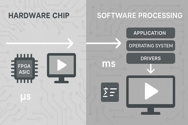
Hardware chips use dedicated circuits, like FPGAs or ASICs, to perform specific tasks like scaling and color correction instantly. Because these operations are hard-wired, they bypass the system’s general processor and operating system, resulting in near-zero latency.
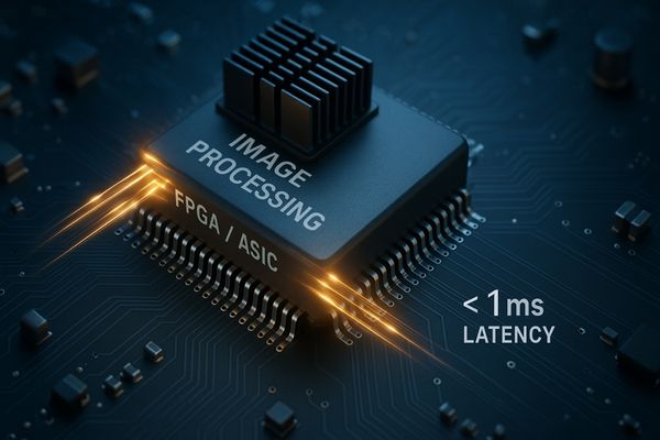
A real-time hardware chip achieves its incredible speed by being purpose-built for a limited set of tasks. Unlike a general-purpose computer processor (CPU) that has to manage an operating system, run multiple applications, and handle various inputs, a dedicated image processing chip does one thing: process video signals as fast as possible. These chips are often Field-Programmable Gate Arrays (FPGAs)4 or Application-Specific Integrated Circuits (ASICs)5. An ASIC is designed from the ground up for a fixed function, offering maximum speed. An FPGA is programmable at the hardware level, offering a balance of speed and some flexibility. When a video signal enters the monitor through an HDMI or SDI port, it goes directly to this chip. The chip’s internal logic is a physical pipeline designed to execute operations like color space conversion, gamma correction, and scaling in parallel. This process happens almost instantaneously, with latency measured in microseconds. This is fundamentally different from software, which must wait for the CPU to be available. This near-zero latency is non-negotiable in surgery, where the surgeon’s hand-eye coordination depends on the display perfectly mirroring their actions. Our MS270P surgical display relies on this principle to provide a fluid, real-time image6.
In what scenarios is software-based image enhancement more flexible than hardware solutions?
A new imaging algorithm is released, but your display’s hardware is now outdated. This forces a costly replacement, but software-based solutions can adapt and evolve with a simple update.
Software is more flexible in scenarios requiring frequent updates, custom workflows, or complex AI-driven analysis. It allows new algorithms for noise reduction, image comparison, or custom display layouts to be deployed without changing the physical monitor hardware.

Software-based image enhancement shines in environments where adaptability is more important than raw speed. This is particularly true in diagnostic imaging7 and clinical review. Because software runs on a general-purpose processor, either within the monitor or on a connected PC, its capabilities can be changed or expanded through code. If a research institution develops a new AI algorithm for detecting microcalcifications8 in mammograms, it can be distributed as a software update to radiologists. This allows a fleet of existing monitors to gain new functionality without any physical hardware changes. Software also enables a high degree of user customization. For example, radiologists can create complex hanging protocols to arrange multiple patient studies across the screen in a specific way, a task managed entirely by software. Our MD45C dual-screen diagnostic monitor uses software to seamlessly manage images across a single large panel, providing the flexibility to display various layouts that a fixed hardware solution could not. While this approach introduces a small amount of latency, it is perfectly acceptable for post-acquisition analysis where real-time feedback is not the primary concern.
| Scenario | Hardware Chip (Best For) | Software Enhancement (Best For) |
|---|---|---|
| Live Surgery | Yes (ultra-low latency) | No (potential for lag) |
| Diagnostic Review | No (too rigid) | Yes (flexible layouts, analysis tools) |
| AI Feature Detection | No (not easily updatable) | Yes (algorithms can be updated) |
| Custom Presets | Limited | Extensive |
Does hardware processing always deliver higher image quality than software?
You might assume hardware is always superior, but some images still lack clarity. This can lead to misinterpretation, because the best quality often comes from a smart combination of hardware and software.
Not necessarily. Hardware excels at the speed and consistent execution of core tasks like color and gamma correction. However, sophisticated software algorithms, especially AI-powered ones, can achieve superior noise reduction, dynamic sharpening, and feature detection that dedicated hardware cannot perform.
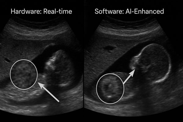
Hardware processing guarantees a high baseline of quality and consistency. It ensures that every frame of a video feed is processed in exactly the same way, delivering stable brightness, accurate color, and adherence to medical standards like DICOM9. This is the foundation of a reliable medical display. However, "higher quality" can also mean more than just consistency. This is where advanced software has the advantage. Software algorithms can perform computationally intensive tasks that are too complex or specific for a general-purpose hardware chip. For example, an AI-powered noise reduction algorithm10 can analyze an image to differentiate between random electronic noise and subtle but important tissue texture. It can then selectively reduce the noise without blurring the critical details. This level of intelligence is difficult and expensive to bake into hardware. The ultimate image quality is often achieved with a hybrid approach. The hardware handles the critical real-time processing to ensure a stable, color-accurate image. Then, advanced software can be applied to provide an additional layer of intelligent refinement. Our 8MP Diagnostic Display11 relies on this principle, using a robust hardware core for clarity and supporting software tools for advanced analytical tasks.
What are the cost implications of hardware vs. software image processing?
You need to upgrade your imaging technology but face a tight budget. Choosing the wrong approach can be expensive in the long run, so understanding the cost structure is crucial.
Hardware processing has a higher upfront cost due to the research, development, and manufacturing of custom chips. Software solutions typically have a lower initial cost but may involve recurring licensing fees, subscription models, or the need for a more powerful host computer.
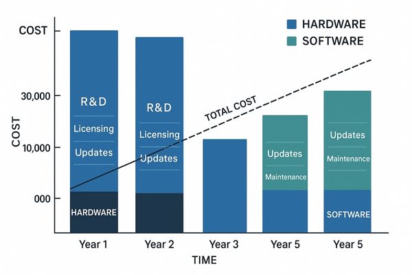
The cost of a medical display is heavily influenced by its image processing architecture12. Developing custom hardware13, especially an ASIC, is a very expensive, front-loaded process that involves lengthy design, prototyping, and fabrication cycles. This investment is reflected in the higher purchase price of a hardware-driven monitor. However, once purchased, the performance is fixed and reliable, with no additional costs for its core function over its lifespan. Software solutions generally have a lower barrier to entry. The development costs can be spread across many users through sales and licensing, leading to a lower initial product price. The potential for ongoing costs is the trade-off. Advanced software features may be sold as optional modules or require an annual subscription for updates and support. Furthermore, demanding software might necessitate a more powerful—and expensive—host computer to run smoothly, which is a hidden cost. Over the total ownership period, the economic comparison can become quite complex. A dependable hardware solution, like the one inside our MD22CA diagnostic monitor, provides excellent long-term value by delivering consistent performance without requiring further investment in software or external computing power.
Cost Structure Comparison
| Cost Factor | Hardware Processing | Software Processing |
|---|---|---|
| Upfront Cost | Higher (custom chip R&D) | Lower (development cost is distributed) |
| Ongoing Costs | Typically None | Potential (subscriptions, licenses) |
| External Hardware | Self-contained | May require powerful host PC |
| Total Lifetime Value | High (predictable, reliable) | Variable (depends on fees and upgrades) |
How does the integration of chips and software benefit surgical displays like those from Reshin?
You need a display that is both fast and intelligent. Choosing a solution focused on only one of these aspects means making a compromise, but a hybrid approach delivers both without drawbacks.
Integrating hardware chips and software allows a display to achieve both ultra-low latency and advanced image enhancement. The chip handles real-time signal processing for surgical precision, while software provides sophisticated color tuning, multi-source viewing modes, and user-defined image presets.
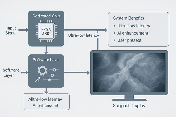
The most advanced surgical displays do not treat hardware and software as an either/or choice. Instead, they integrate them to create a system that is greater than the sum of its parts. This hybrid model leverages the core strength of each technology. At the heart of our surgical monitors14 is a dedicated hardware processing chip. This component is solely responsible for taking the raw video feed from an endoscope or surgical camera and putting it on the screen with the lowest possible latency. This guarantees the immediate visual feedback that surgeons require for precise, confident movements. Layered on top of this hardware foundation is our sophisticated software and firmware. This software does not interfere with the real-time video path. Instead, it acts as a control layer, giving the surgeon powerful tools to optimize the image for their specific needs. For example, the software allows a surgeon to instantly switch between custom look-up tables (LUTs)15 that are pre-calibrated to enhance visuals for different types of surgery, like laparoscopy or urology. The software also manages features like picture-in-picture (PIP) modes16 to display a secondary source, or onscreen menus for adjusting brightness and contrast. This integrated design, perfected in our MS321PC 4K monitor, delivers the best of both worlds: the uncompromised speed of hardware and the intelligent flexibility of software.
Conclusion
Real-time chips provide the speed essential for surgery, while software offers the flexibility for advanced analysis. The best systems integrate both, delivering fast, reliable, and intelligently enhanced images for modern medicine. To explore medical displays with integrated real-time hardware and intelligent software solutions, contact Reshin at martin@reshinmonitors.com.
- Explore this link to understand the cutting-edge innovations that are shaping the future of medical displays, enhancing diagnostics and patient care. ↩
- Learn about hardware-based processing to see how it contributes to display performance and stability in medical environments. ↩
- Discover how software-based enhancements can adapt and analyze data, making displays more versatile and effective in clinical settings. ↩
- Explore this link to understand how FPGAs work and their significance in real-time processing applications. ↩
- Learn about ASICs and how they are optimized for specific tasks, enhancing performance in various technologies. ↩
- Discover the technology behind real-time image processing in surgical displays and its critical role in medical procedures. ↩
- Exploring advancements in diagnostic imaging can provide insights into how technology is evolving to improve patient care and outcomes. ↩
- Understanding AI’s role in detecting microcalcifications can highlight its impact on early cancer detection and treatment strategies. ↩
- Discover the significance of DICOM standards in ensuring quality and consistency in medical imaging. ↩
- Explore this link to understand how AI-powered noise reduction enhances image quality, especially in medical imaging. ↩
- Learn about the advanced features of the 8MP Diagnostic Display and how it improves medical imaging accuracy. ↩
- Understanding image processing architecture can help you make informed decisions about medical display technologies and their costs. ↩
- Exploring the benefits of custom hardware can provide insights into its long-term value and performance in medical applications. ↩
- Explore how surgical monitors enhance precision and efficiency in surgeries, providing critical visual feedback for surgeons. ↩
- Learn about the role of LUTs in optimizing surgical visuals for different procedures, enhancing clarity and detail. ↩
- Discover how PIP modes can improve surgical workflows by allowing simultaneous viewing of multiple video sources. ↩