Investing in a high-end monitor without understanding its core compliance features is a risk. This can lead to inconsistent diagnoses, non-compliance during audits, and wasted capital on equipment that fails in practice.
A DICOM-compliant display lives in GSDF, sustained luminance (CBS+ALC), right resolution (dual 5MP vs single 12MP), uniformity, and per-window control—protecting accuracy in PACS and mammography.
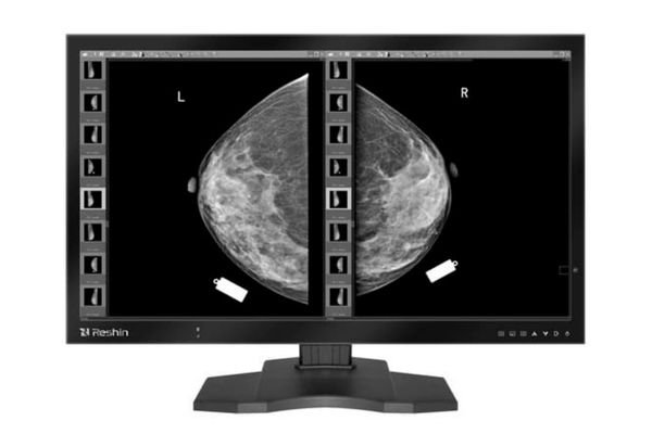
A DICOM-compliant display must pass GSDF acceptance1 and prove constancy via scheduled QC; otherwise fleets drift and audits fail. This guide explains how consistent grayscale rendering, stable luminance, appropriate resolution, panel uniformity, and smart workflow tools work together to protect the integrity of every read, reducing diagnostic uncertainty2 and improving clinical efficiency. Peak nits don’t guarantee diagnostic constancy—verified GSDF and sustained luminance do.
DICOM GSDF & JND consistency: acceptance and constancy QC (DICOM-compliant display)
Buying a "DICOM-calibrated" monitor is not enough. Without ongoing checks, its grayscale performance can drift, making it impossible to guarantee consistent interpretations across different workstations or over time.
A display must pass initial GSDF acceptance and then prove constancy with scheduled automated QC. This ensures Just Noticeable Difference (JND) steps remain perceptually uniform, keeping fleets aligned.
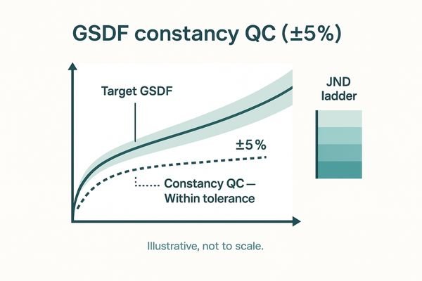
A display’s core diagnostic responsibility is to render grayscales according to the DICOM Part 14 Grayscale Standard Display Function (GSDF)3. Constancy checks keep GSDF deviation within ±5%, preserving JND steps across time and sites. True compliance requires an initial acceptance test followed by routine constancy QC4 to prevent performance drift. The monitor must actively participate in its own QC. A diagnostic display like the MD33G includes built-in DICOM-QC and PACS-QC luminance tracking for scheduled self-tests. This embedded approach logs performance and flags deviations, ensuring every display in a multi-site PACS network provides a consistent, reliable view for radiologists (±5% GSDF tolerance).
Calibrated luminance & stability (CBS) with ambient light compensation (ALC)
A radiologist’s reading environment is not a static lab. Changing ambient light throughout the day can wash out screen contrast and cause eye strain, compromising diagnostic accuracy.
Trusted performance comes from sustained, verified luminance under real-world conditions. CBS counters backlight aging, and ALC adapts to room light, ensuring year-two images perceptually match day-one.
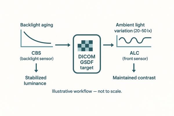
The luminance value that matters is the stable, calibrated brightness delivered under real-world reading conditions. A Constant Brightness System (CBS)5 uses an integrated sensor to counteract backlight aging. An Ambient Light Compensation (ALC)6 sensor faces the user, measuring room light to maintain display performance. ALC targets a consistent contrast ratio across typical reading-room illuminance (e.g., ~20–50 lx), preventing image washout and reducing eye strain. This functionality is available on reliable clinical review displays like the MD46C. Together, CBS+ALC fulfill the core promise of DICOM compliance: day-2-year images match day-1.
Resolution & pixel pitch for reading: dual 5MP vs single 12MP
Choosing between a dual 5MP setup and a single 12MP display can be confusing. Sticking with legacy setups may seem safe, but it can create workflow bottlenecks and ergonomic issues.
Two matched 5MP monitors are a robust choice, but a single 12MP display can consolidate views and speed up comparisons. The decision depends on workload, with mammography gaining most from 12MP.
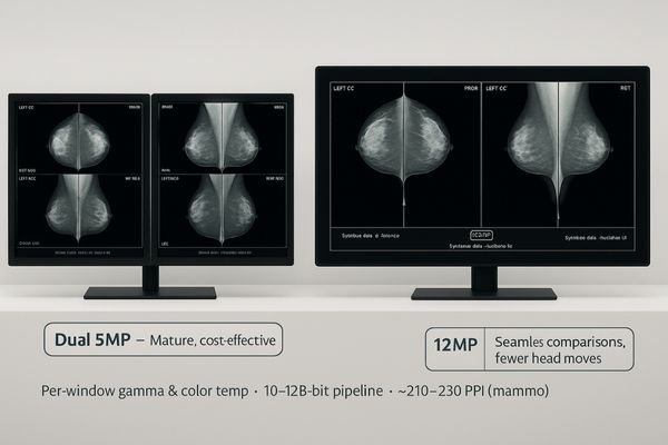
The debate between dual 5MP and single 12MP displays comes down to workflow and ergonomics. Mammography targets ~210–230 PPI; general PACS ~100–150 PPI; higher PPI improves micro-calc visibility alongside 10–12-bit pipelines. While a dual 5MP setup is cost-effective, a single 12MP display like the MD120C offers a seamless canvas ideal for mammography comparisons, reducing eye travel. The key is per-window control over gamma and color temperature. Centers with heavy comparison needs see the greatest benefit from 12MP consolidation.
| Setup | Pros | Watch-outs | Best for |
|---|---|---|---|
| Dual 5MP | Mature workflow, cost-effective | Bezel breaks comparison, more eye travel | Mixed PACS |
| Single 12MP | Seamless comparisons, fewer head moves | Needs per-window gamma/CTemp | Mammography, prior-heavy reads |
Uniformity, black level & noise: protecting subtle lesions and micro-calcifications
Luminance non-uniformity or a high black level can mask faint pathologies, especially at the edges of the screen. This is a subtle but critical failure that undermines diagnostic confidence.
Subtle findings fail first at the edges. Uniformity compensation, low black levels, and high native contrast are essential to preserve the conspicuity of faint lesions and micro-calcifications.
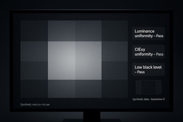
Resolution alone does not guarantee diagnostic accuracy. Luminance uniformity is just as critical. Uniformity compensation corrects center-to-corner luminance/CIExy variance, protecting lesion conspicuity at the periphery. A low black level, which enables a high native contrast ratio, is equally important for visualizing differences in dark tissues. Minimizing panel noise ensures the image is not obscured by electronic artifacts. A high-performance 5MP grayscale mammography monitor7 like the MD52G is engineered to excel in these areas, giving radiologists the confidence to make decisions on borderline cases.
Workflow tools: per-window control, multiview, auto-rotation & QC on OSD
A powerful display with a clumsy interface is a frustrating tool. Radiologists lose valuable time navigating complex menus or manually adjusting settings for every study.
Multiview is only useful when each window locks its own parameters and layouts are on the OSD. Smart tools like auto-rotation and built-in QC turn daily checks into seconds.
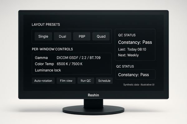
The most effective diagnostic displays8 enhance workflow with smart, time-saving tools. Their value is unlocked with per-window gamma, color temperature, and luminance lock, plus OSD hotkeys for layout toggles and one-touch film-view. A fusion display like the MD85CA allows a radiologist to lock settings for each image source, ensuring accurate comparison. Other enhancers include auto-rotation for portrait-oriented images and OSD-based QC for quick checks without launching separate software. Each tool is designed to reduce clicks and "micro-steps," compounding into significant throughput gains across a busy department.
Conclusion
Ensure your DICOM-compliant display provides sustained compliance through automated QC, stable luminance, appropriate resolution, panel uniformity, and intuitive workflow tools. Get a GSDF acceptance template and a 12MP/dual-5MP selection sheet to accelerate your next deployment. ✅
👉 For expert guidance and tailored Reshin monitor solutions, contact martin@reshinmonitors.com today.
-
Understanding GSDF acceptance is crucial for ensuring accurate medical imaging and compliance with standards. ↩
-
Exploring ways to reduce diagnostic uncertainty can enhance clinical outcomes and improve patient care. ↩
-
Understanding GSDF is crucial for ensuring accurate grayscale rendering in medical imaging, enhancing diagnostic reliability. ↩
-
Exploring routine constancy QC will help you grasp its importance in maintaining display performance and image quality over time. ↩
-
Understanding CBS can enhance your knowledge of display technology and its impact on image quality. ↩
-
Exploring ALC will reveal its crucial role in maintaining optimal viewing conditions and reducing eye strain. ↩
-
Exploring the advantages of high-performance monitors can enhance your knowledge of their impact on diagnostic accuracy and patient care. ↩
-
Explore how diagnostic displays can streamline workflows and improve efficiency in medical imaging. ↩


