How to Plan Multi-Unit Diagnostic Display Deployment Without Future Replacement Chaos

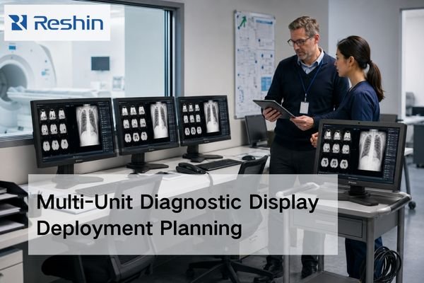
Radiology team reviewing a multi-unit diagnostic display deployment with matched medical monitors in a PACS reading room

Effective multi-unit diagnostic display deployment is a lifecycle planning exercise, not a simple purchase. To avoid future replacement chaos, planning should start with a clear baseline, a realistic matching rule across workstations, a practical QA path, and replacement logic that is defined before expansion begins. A capable diagnostic monitor manufacturer should be able to support that full deployment logic, not just deliver the first batch.

What Really Matters in OR Display Integration

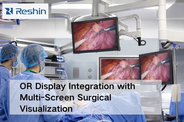
Integrated operating room with multiple surgical displays showing consistent endoscopic images during OR display integration

Successful OR display integration depends more on system-level predictability, stable signal switching, practical mounting, and multi-unit consistency than on the standalone specifications of a single monitor. A capable surgical monitor manufacturer should help the project stay controllable across the full display layer, not just offer a stronger spec sheet.

What is PACS and how does it affect radiology monitor workflows?

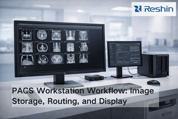
Bright radiology IT workspace showing a PACS workstation monitor beside network storage and a switch, illustrating image distribution to radiology workflows

PACS (Picture Archiving and Communication System) is the image backbone that stores, organizes, and delivers studies across radiology. It shapes monitor workflows through PACS viewers and hanging protocols—how images are laid out, rendered, windowed, and compared with priors—so diagnostic display performance must be validated inside real PACS workflows, not in isolation.

What should you watch for when using multi-stream transport for a dual-screen imaging workstation?

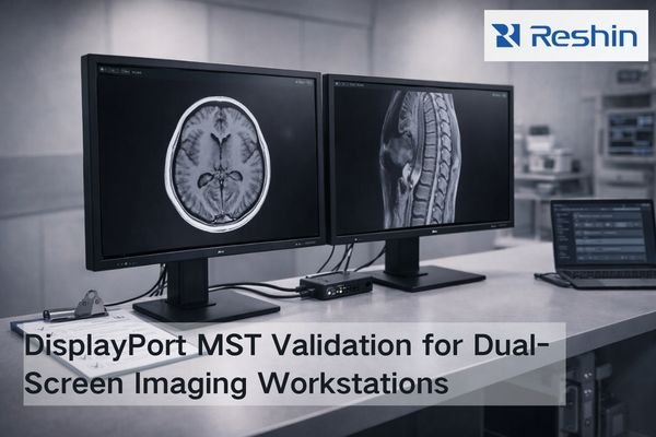
Dual-screen imaging workstation with DisplayPort MST hub and two medical-grade monitors showing grayscale images for stable mode validation

Multi-Stream Transport (MST) allows one DisplayPort output to drive multiple displays through daisy-chaining or hubs, simplifying cabling for dual-screen imaging workstations while introducing extra negotiation steps and variables in the signal chain. Success depends on bandwidth management, stable EDID handling, consistent display enumeration, and validated mode sets that prevent silent downgrades affecting image quality and workflow consistency in demanding clinical environments.

Do Medical Displays Still Need Composite/S-Video Inputs?

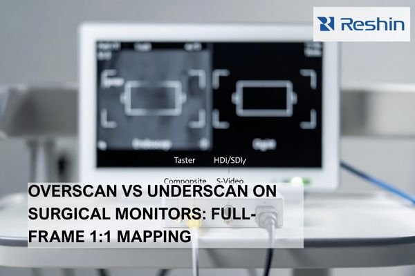
Medical display with Composite and S-Video connectors beside a digital cable, illustrating legacy analog inputs and converter-based transition in OR video routing

Most modern OR workflows don’t need Composite/S-Video on every medical display. However, long-lifecycle legacy devices can still justify analog support at specific endpoints or through converters. The decision should be inventory-driven: identify which sources are still analog, estimate the downtime impact if they fail, and standardize on digital while keeping a validated fallback for critical legacy feeds.

How Should Ambulatory Surgery Centers Choose Surgical Monitors to Reduce Maintenance?

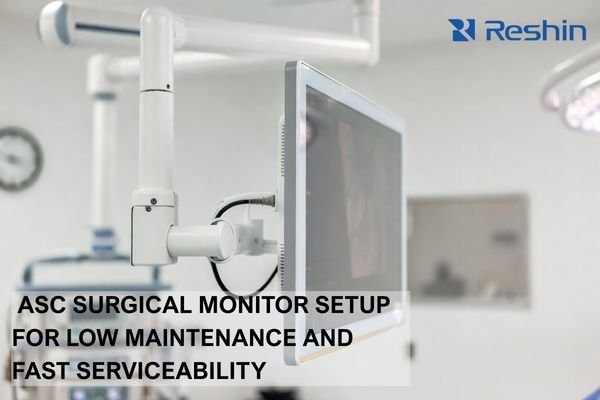
Boom-mounted ASC surgical monitor with sealed cleanable front and strain-relieved cables for reduced maintenance and downtime

Ambulatory Surgery Centers should choose surgical monitors by prioritizing predictable signal behavior, OR-ready cleanability with sealed housings, robust mounting with strain relief, and fast serviceability. In ASC environments, time-to-restore is the critical KPI: small issues like intermittent video, connector looseness, or cleaning-related wear can quickly cascade into delayed cases, rescheduled lists, and lost revenue.

What Requirements Does Intraoperative 3D Imaging (3D Fluoro/CT) Put on Displays?

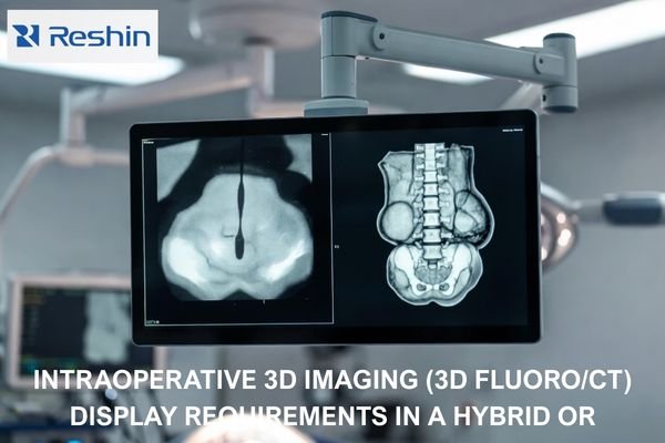
Hybrid OR medical-grade display showing live 2D fluoroscopy and intraoperative 3D cone-beam CT volume side by side

Intraoperative 3D imaging (3D fluoro/CT) needs displays that keep low-contrast cues visible for navigation, render fine detail without added artifacts during interaction, and stay consistent across sources and viewing positions. Priorities include stable grayscale/brightness behavior, restrained processing (no halos or over-sharpening), reliable tone mapping, and fast, predictable source switching.

Why Does Otologic Microsurgery Rely More on Low-Latency Surgical Monitors?

Reshin 4K surgical monitor displaying high-resolution endoscopic image of otologic microsurgery

Otologic microsurgery relies more on low‑latency surgical monitors because even minimal delays of 50–100 milliseconds can cause surgical instruments to overshoot intended movements during high‑magnification operations within tight anatomical spaces. These procedures require instantaneous visual–motor synchronization, and latency disrupts precision, reduces surgeon confidence, and increases the risk of unintended trauma to delicate structures such as ossicles, cochlea, and facial nerve pathways.

How Should You Choose a Surgical Monitor for Neurosurgery Microscope Video Output?

Neurosurgery OR scene with a surgical microscope and an articulated surgical monitor displaying a non-graphic microscope-view image with fine microvascular detail.

Choosing surgical monitors for neurosurgery microscope video output requires preserving micro-detail and maintaining repeatability across real OR signal chains rather than pursuing headline specifications. Prioritize clean scaling with minimal artifacts, confirm delivered formats and latency behavior, and lock stable brightness and picture modes that prevent drift during long cases to support both surgical precision and consistent team communication.