How to Evaluate Grayscale and Luminance Consistency for Diagnostic Displays

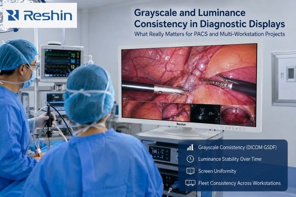
Operating room scene with surgeons viewing a large medical display illustrating grayscale and luminance consistency in diagnostic imaging

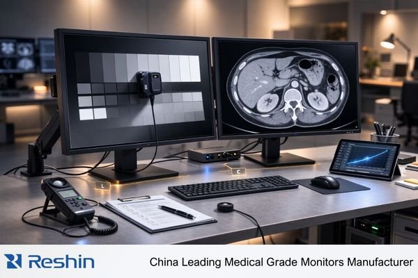
Effective evaluation of diagnostic displays goes beyond headline specs. It requires checking grayscale consistency through DICOM GSDF behavior, separating luminance into long-term stability and screen uniformity, and confirming that those conditions can stay close enough across multiple workstations. For projects that depend on controlled PACS reading conditions, working with a dedicated PACS monitor manufacturer is usually a more useful starting point than comparing isolated specs alone.

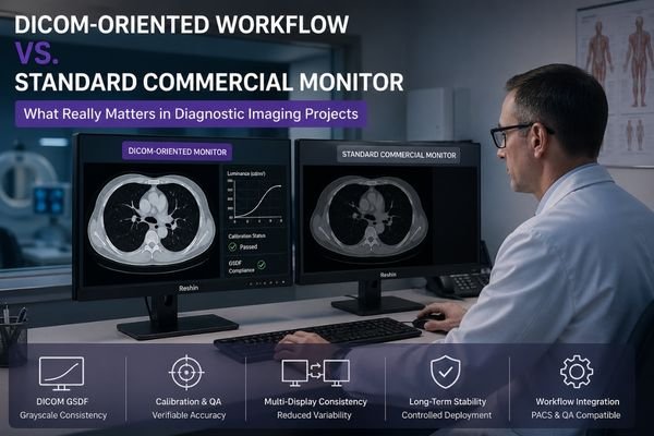
DICOM-Oriented Workflow vs Standard Commercial Monitor: What Buyers Should Know

Radiologist comparing a DICOM-oriented diagnostic monitor with a standard commercial monitor in a medical imaging workstation

A DICOM-oriented workflow is built around stable grayscale presentation, repeatable calibration logic, and controlled reading conditions over time. A standard commercial monitor may display a medical image, but it is usually not designed to support the grayscale behavior, QA discipline, and workflow control that a diagnostic process depends on. A dedicated diagnostic monitor manufacturer is typically working toward a different project goal than a standard commercial display vendor.

What is PACS and how does it affect radiology monitor workflows?

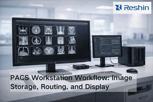
Bright radiology IT workspace showing a PACS workstation monitor beside network storage and a switch, illustrating image distribution to radiology workflows

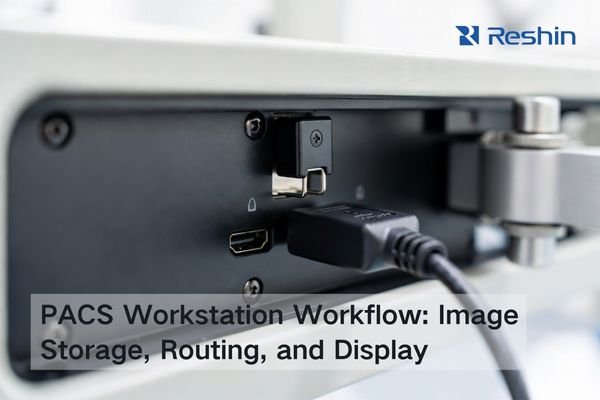
PACS (Picture Archiving and Communication System) is the image backbone that stores, organizes, and delivers studies across radiology. It shapes monitor workflows through PACS viewers and hanging protocols—how images are laid out, rendered, windowed, and compared with priors—so diagnostic display performance must be validated inside real PACS workflows, not in isolation.

How do you verify auto-rotation and portrait lock on a diagnostic monitor?

Radiology workstation with two diagnostic monitors in portrait and landscape modes showing grayscale images for auto-rotation and portrait lock testing

To properly verify auto-rotation and portrait lock on diagnostic monitors, test both features across multiple scenarios including system restarts, sleep/wake cycles, and multi-display setups. Check for consistent performance, proper orientation detection, and reliable locking over extended periods.

A Brief Introduction to Medical Grade Monitors?

Medical-grade monitor beside a consumer display in a clinical room, showing consistent DICOM-style grayscale and a non-graphic endoscopy inset

A medical-grade monitors is a specialized display built for clinical environments. It shows images from medical equipment like CT scanners or endoscopes with high accuracy and stability. This is essential for applications like surgery and radiology diagnosis, ensuring doctors see precise information to make critical decisions.

Do Medical Displays Still Need Composite/S-Video Inputs?

Medical display with Composite and S-Video connectors beside a digital cable, illustrating legacy analog inputs and converter-based transition in OR video routing

Most modern OR workflows don’t need Composite/S-Video on every medical display. However, long-lifecycle legacy devices can still justify analog support at specific endpoints or through converters. The decision should be inventory-driven: identify which sources are still analog, estimate the downtime impact if they fail, and standardize on digital while keeping a validated fallback for critical legacy feeds.

How Should You Set Overscan/Underscan on Surgical Monitors?

Surgical monitor showing overscan vs underscan with cropped edge UI on one side and full-frame 1:1 pixel mapping on the other

For most OR video chains, the safest default is underscan / 1:1 pixel mapping so the full frame remains visible, including edge UI such as scale bars, measurement markers, patient identifiers, and warning banners. Use overscan only as a last resort to hide unavoidable edge artifacts that cannot be corrected upstream, and keep it minimal because scaling can soften detail, alter geometry, and reduce consistency across side-by-side surgical monitors.