Inconsistent medical images create diagnostic uncertainty. A subtle change in brightness or color can hide a critical finding. To prevent this, strict imaging standards ensure that displays show accurate and reliable information for every patient, every time.
This article explains the essential imaging standards for medical displays. We will explore the framework of these rules, covering DICOM for image consistency, IEC and FDA for safety, calibration for accuracy, and how guidelines are evolving for future technologies.
These standards are not just technical rules; they are a promise of quality and safety in patient care. Understanding them is key to choosing the right equipment. Let us now examine how this system of standards works to ensure diagnostic confidence.
DICOM Compliance Ensures Consistent Grayscale Display
An X-ray might look different on two separate screens. This variation can cause a doctor to miss a problem. The DICOM standard solves this by making sure both screens show the exact same image.
DICOM is the universal language for medical images. Its grayscale rules ensure that any digital image value is shown with a specific, uniform brightness. This consistency is the foundation of accurate radiological diagnosis.
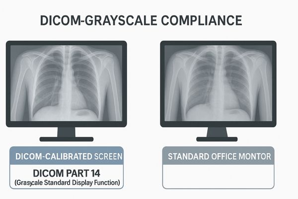
DICOM, which stands for Digital Imaging and Communications in Medicine, provides a rulebook for medical images. The most important rule for displays is in DICOM Part 14. It defines the Grayscale Standard Display Function (GSDF)1. Think of the GSDF as a universal translator for brightness. It ensures that a specific pixel value in a CT or X-ray image always corresponds to the same level of light output on any compliant monitor. This is crucial because the human eye does not perceive changes in brightness linearly. The GSDF is specifically tuned to human vision, making it easier for radiologists to spot subtle differences in dark and light areas. This means a faint shadow suggesting a tumor will be equally visible on any DICOM-compliant display. Our MD26GA – 2MP Grayscale Diagnostic Monitor is built to strictly follow this DICOM Part 142 standard, providing the dependable grayscale performance needed for primary diagnosis.
IEC and FDA Standards Define Safety and Performance
A display that is not certified can be an electrical risk in a hospital. This creates a danger for both patients and staff. Official standards from bodies like the IEC and FDA exist to guarantee every device is safe.
A complex hierarchy of standards governs medical displays. Global bodies like the IEC set the baseline for safety, while national agencies like the FDA add specific performance and regulatory requirements for market approval.
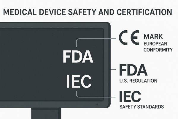
Medical displays are not like consumer electronics; they are highly regulated medical devices. They must pass a series of checks defined by a clear structure of standards. At the top are global standards from the International Electrotechnical Commission (IEC). The IEC 60601-13 standard is the primary benchmark for the safety of all medical electrical equipment, covering risks like electrical shock and fire. Think of this as the international safety passport. Below this global standard, national or regional regulatory bodies add their own rules. In the United States, the Food and Drug Administration (FDA) requires a 510(k) submission4 to prove a device is safe and effective. In Europe, a CE mark is required. The MD50C – 5MP Color Mammography Monitor must meet these stringent requirements, including the FDA’s specific rules under the Mammography Quality Standards Act (MQSA), to be used for breast cancer screening.
| Standards Body | Scope | Key Function | Example Standard |
|---|---|---|---|
| IEC | Global | Sets baseline electrical and mechanical safety | IEC 60601-1 |
| FDA | National (USA) | Regulates market approval for safety & efficacy | 510(k) Premarket Notification |
| CE Mark | Regional (Europe) | Certifies compliance with EU safety directives | Medical Device Regulation (MDR) |
Calibration Protocols Maintain Long-Term Display Accuracy
Over time, every monitor’s performance fades. Its brightness and color accuracy can slowly change. This drift can make important diagnostic details less visible, so regular calibration is necessary to restore its precision.
All displays age, and their visual performance changes. Routine calibration is a vital maintenance process that measures the display’s output and readjusts it. This ensures its brightness and color remain accurate for reliable diagnosis.
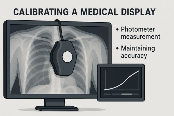
A medical display is a precision instrument, and like any instrument, it needs regular tuning to stay accurate. From the moment it is first used, its backlight system begins to age, causing a gradual decrease in brightness and shifts in color. This is not a defect; it is a normal physical process. If left uncorrected, this performance drift could compromise diagnostic accuracy. Calibration5 solves this problem. The process uses a highly sensitive light-measuring device, called a photometer6, to analyze the screen’s actual light and color output. Special software compares these real-world measurements against the required standard, such as DICOM GSDF or a specific color profile. The software then creates a new correction map inside the monitor to restore its performance to the original factory standard. The MD51CHY – 34" 5MP Diagnostic Monitor for X-ray Imaging supports advanced calibration to ensure its wide-format screen provides consistent, accurate images for its entire operational life.
Imaging Standards Support Cross-Platform Consistency
A doctor in another city needs to review a patient’s scan. They must trust that the image they see is identical to the original. Shared imaging standards build a reliable network for this kind of collaboration.
Standardized imaging creates a trusted visual language across all devices and locations. This allows clinicians to share images between different hospitals with confidence, knowing a diagnosis made on one compliant display is valid on another.
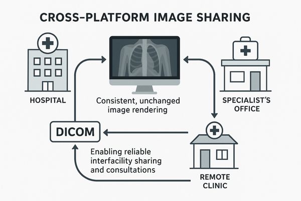
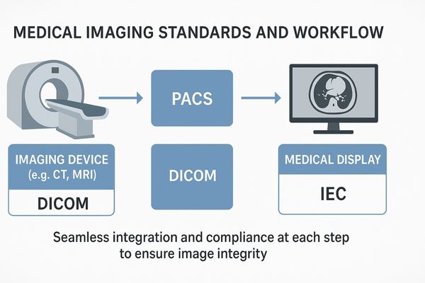
Modern medicine relies heavily on teamwork, often across different buildings or even countries. Teleradiology allows a specialist to read a scan from a rural clinic, while a tumor board may bring together doctors from multiple hospitals to review a complex case. This collaboration is only possible because of universal standards. The DICOM standard7 ensures the entire imaging chain, from the scanner that captures the image to the PACS server that stores it and the display that shows it, speaks the same language. This guarantees that the image data is never altered and is rendered correctly at every point. It means a radiologist performing a remote consultation sees the exact same image as the doctor beside the patient. Our MD46C – Dual-screen Diagnostic Monitor (Single Panel) is designed for these workflows. Its single-panel construction eliminates any visual gap or inconsistency between two images, which is perfect for comparing a current scan to a previous one from another facility.
Evolving Guidelines Reflect Advances in Medical Imaging
New technologies like digital pathology and AI are introducing new visual information. Old standards focused on grayscale were not designed for this. Therefore, the guidelines must evolve to keep pace with these innovations.
Medical imaging technology is advancing quickly. As new tools like color pathology images and AI overlays become common, standards bodies are updating guidelines to ensure this new visual data is displayed accurately and safely.
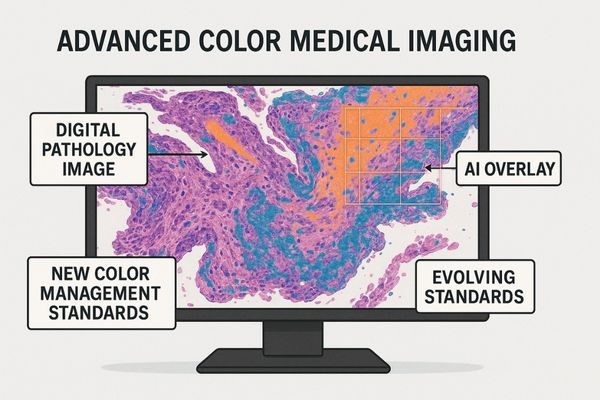
The world of medical imaging is expanding beyond traditional grayscale X-rays. Fields like digital pathology and dermatology rely on precise color to identify tissue types and disease markers. For these applications, DICOM GSDF8 is not enough. Here, color standards like those defined by the International Commission on Illumination (CIE)9 become important. These standards help ensure that the color a pathologist sees on the screen is a true match to the color seen through a microscope. Furthermore, new techniques fuse images from different machines, like overlaying a color PET scan on a grayscale CT scan. Displays must manage both color and grayscale data at once. AI is also adding new layers of information. As these technologies mature, standards are being updated to include new color profiles and rules for handling overlays. The MD22CA – 2MP Color Diagnostic Monitor is built for this modern environment, offering the calibrated color accuracy needed for these evolving clinical applications.
Conclusion
Imaging standards provide the foundation of trust in medical diagnosis. They ensure every display delivers the safety, accuracy, consistency, and future-readiness required for the highest level of patient care.
📩 Looking to equip your facility with displays that meet the latest imaging standards? Reach out to Martin at martin@reshinmonitors.com for tailored solutions from Reshin.
-
Understanding GSDF is essential for ensuring accurate medical imaging and diagnosis, making this resource invaluable for professionals. ↩
-
Exploring DICOM Part 14 guidelines will enhance your knowledge of medical imaging standards, crucial for quality healthcare. ↩
-
Understanding the IEC 60601-1 standard is crucial for ensuring the safety and effectiveness of medical devices. ↩
-
Exploring the 510(k) submission process will provide insights into how medical devices are evaluated for safety and efficacy. ↩
-
Understanding calibration is crucial for maintaining diagnostic accuracy in medical displays, ensuring reliable imaging. ↩
-
Exploring how a photometer functions can enhance your knowledge of the calibration process, vital for accurate medical imaging. ↩
-
Understanding the DICOM standard is crucial for anyone involved in medical imaging, as it ensures compatibility and quality across different systems. ↩
-
Understanding DICOM GSDF is crucial for grasping how color standards impact medical imaging accuracy. ↩
-
Exploring CIE’s role will enhance your knowledge of color accuracy in medical diagnostics. ↩