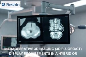
Radiologists depend on clear images for vital diagnoses. But what makes a monitor truly "medical-grade"? Is your display showing the full picture?
For radiology monitors, the most crucial image quality parameters are exceptional grayscale depth, appropriate luminance and contrast ratio for the specific viewing environment, sufficient pixel density tailored to the imaging modality, outstanding screen uniformity, and fast response times for dynamic studies. These ensure diagnostic accuracy.

Understanding these technical details isn’t just for engineers like myself; it’s fundamental for every healthcare professional involved in image-based diagnosis. Each parameter plays a distinct role in how clearly and accurately a patient’s condition can be assessed. Let’s delve into why these specific characteristics are so critical in the demanding field of radiology.
How does grayscale display capability impact the diagnostic accuracy of radiology monitors?
In a world full of color, radiology thrives on shades of gray. But not all grays are created equal. How much difference does it really make?
Grayscale display capability, particularly the bit depth (e.g., 12-bit to 16-bit, offering thousands of gray shades), is paramount. It allows radiologists to distinguish subtle differences in tissue density, which is critical for identifying pathologies like faint nodules or early infiltrates.

In my years at Reshin, I’ve seen firsthand that grayscale depth is the absolute core of diagnostic imaging. Radiological images, whether X-rays, CT scans, or MRIs, rely almost entirely on subtle differences in grayscale to differentiate between various tissues and identify abnormalities. Standard consumer monitors typically display 8-bit grayscale, which gives you about 256 shades. Medical-grade radiology monitors, however, often work with 10-bit, 12-bit, or even higher (internally processing 14-bit or 16-bit data), translating to 1,024 to over 4,096 distinct shades of gray.
I recall a situation where a partner hospital was experiencing a higher-than-average rate of missed early-stage lung nodules. When we investigated, one contributing factor was their use of older 10-bit monitors that weren’t optimally calibrated for the subtle transitions needed. Upgrading to newer displays with true 12-bit output and better DICOM calibration capabilities made a noticeable difference. The ability to perceive these minute density variations is especially critical in applications like early cancer screening. It’s these tiny, almost imperceptible differences that can signal a developing issue.
Grayscale Bit Depth Comparison
| Bit Depth | Shades of Gray | Typical Use Case |
|---|---|---|
| 8-bit | 256 | Consumer displays |
| 10-bit | 1,024 | Basic medical review |
| 12-bit | 4,096 | Diagnostic radiology |
| 14-bit+ | 16,384+ | Advanced imaging (e.g., mammography internal processing) |
This increased depth is what allows our Reshin displays to reveal the nuances that are essential for confident diagnosis.
What role do maximum brightness and contrast ratio play in distinguishing tissue densities on radiology monitors?
Ever tried reading a dim screen in a bright room? It’s frustrating. For radiologists, it can mean missing something vital. So how bright is bright enough?
Maximum brightness (luminance) and a high contrast ratio are crucial. Sufficient brightness allows clear image viewing even in well-lit environments, while a high contrast ratio enables the differentiation of subtle variations in tissue density, making abnormalities more conspicuous.

Brightness, or luminance, and contrast ratio aren’t just about making images look good; they must be tailored to the clinical scenario. I’ve learned that a one-size-fits-all approach doesn’t work. For typical PACS (Picture Archiving and Communication System) workstations in a controlled, dimly lit reading room, a maximum luminance of around 300 to 500 cd/m² (candelas per square meter) and a contrast ratio of 1000:1 or higher is generally sufficient. This provides clear, detailed images without causing eye strain during long reading sessions.
However, the demands change dramatically in other environments. For instance, in an operating room during a fluoroscopy-guided procedure, ambient lighting is often much brighter. Here, a monitor needs to achieve a much higher luminance, often exceeding 1000 cd/m², to ensure that critical details in an angiogram, for example, are not washed out. I once consulted on a case where a monitor with unoptimized dynamic contrast, struggling in a bright OR, contributed to a misjudgment of vascular stenosis during a stenting procedure. It highlighted for me just how vital scenario-specific parameter tuning is. The monitor must adapt, not the diagnostician.
Brightness & Contrast Needs by Environment
| Environment | Typical Luminance (cd/m²) | Minimum Contrast Ratio | Key Challenge |
|---|---|---|---|
| PACS Reading Room | 300 – 500 | 1000:1 | Sustained viewing comfort |
| Operating Room (OR) | >1000 | 1500:1+ | High ambient light |
| Mammography | 420 – 700+ (DICOM GSDF) | 1200:1 | Subtle microcalcifications |
At Reshin, we ensure our displays offer a wide dynamic range and stable luminance to meet these varied demands.
How does pixel density (PPI) affect the clarity of radiological images on monitors?
We hear a lot about "high resolution" in consumer tech. But in radiology, it’s about seeing the tiniest details. How many pixels are truly needed?
Higher pixel density (Pixels Per Inch, or PPI) directly translates to sharper, more detailed radiological images. This is critical for visualizing fine structures, such as microcalcifications in mammography or subtle trabecular bone patterns, which can be lost on lower-resolution displays.

The required resolution and pixel density are very much dependent on the imaging modality. It’s not a simple case of "more is always better," although for certain applications, it truly is. For general radiology, like viewing chest DR (Digital Radiography) images, a 2-megapixel (MP) or 3MP monitor might suffice, providing adequate detail. However, when you move to mammography, the requirements skyrocket. To visualize tiny microcalcifications, which can be as small as 0.1mm and are critical early indicators of breast cancer, you absolutely need 5MP or even higher resolution displays (often 8MP or 12MP pairs).
I remember an instance where a clinic using older monitors with a pixel density around 100 PPI for mammography review struggled with differentiating microcalcification clusters from benign vascular shadows. This led to unnecessary biopsies and patient anxiety. Once they upgraded to specialized mammography displays with much higher PPI, the diagnostic confidence improved significantly. Furthermore, with the rise of multi-modality image fusion, like PET-CT, higher resolution is crucial to prevent artifacts or loss of detail in the fused regions, which could otherwise compromise the diagnostic interpretation.
Recommended Resolution by Modality
| Modality | Minimum Resolution | Typical PPI Range | Critical Detail Example |
|---|---|---|---|
| Chest DR/CR | 2MP – 3MP | 90 – 110 | Lung nodules, faint infiltrates |
| CT/MRI (General) | 2MP – 4MP | 100 – 120 | Soft tissue structures, lesions |
| Mammography | 5MP – 12MP (pair) | 170 – 210+ | Microcalcifications (0.1-0.3mm) |
| PET-CT Fusion | 3MP – 5MP | 110 – 130 | Accurate overlay of functional/anatomical data |
Reshin focuses on providing modality-specific displays because we understand these nuanced requirements.
Why is screen uniformity critical for consistent interpretation of medical images on radiology monitors?
Imagine reading a book where the ink fades at the edges. Frustrating, right? For a radiologist, a non-uniform screen can lead to diagnostic errors.
Screen uniformity, meaning consistent brightness and color across the entire display surface, is vital. Non-uniformity can cause a lesion to appear different depending on its position on the screen, potentially leading to misinterpretation of its density or significance.

Screen uniformity is one of those parameters that might not sound exciting, but its impact on diagnostic accuracy is profound. If there’s a significant difference in brightness between the center and the edges of a monitor – say, greater than 10% – a radiologist might misjudge the density of a lesion. For example, a liver hemangioma might appear more like a cyst if viewed in a dimmer portion of the screen. The DICOM Part 14 Grayscale Standard Display Function (GSDF) recommends that luminance uniformity variations should be within a certain percentage, often striving for less than 5% across the usable screen area after calibration.
I recall a case where a radiology department was using monitors that, after a year of use, developed about a 20% brightness loss at the edges compared to the center. This deviation from uniformity contributed to two missed subtle lesions on abdominal CT scans, which were later caught on review with a properly uniform display. This highlights why robust backlight technology, like direct-lit LED backlights, often offers better foundational uniformity and stability over time compared to some edge-lit designs, especially in larger displays. Regular quality assurance and calibration are key to maintaining this uniformity.
Uniformity Impact
| Uniformity Level | Potential Issue | Recommendation |
|---|---|---|
| Poor (>15% variance) | High risk of misinterpreting lesion density/visibility | Replace or repair |
| Fair (10-15% variance) | Possible impact on subtle findings | Monitor closely, calibrate |
| Good (<10% variance) | Generally acceptable for review | Regular QA |
| Excellent (<5% variance) | Ideal for primary diagnosis | Maintain via calibration |
At Reshin, we use stringent quality control for luminance uniformity to prevent such diagnostic pitfalls.
Conclusion
Key parameters like grayscale, brightness, contrast, pixel density, and uniformity are non-negotiable for radiology monitors. Prioritizing these ensures diagnostic accuracy and patient safety.


