Choosing a 4K display for the OR based on consumer specs can be a critical mistake. This approach often leads to poor visibility under surgical lights and unreliable performance mid-procedure.
- Reshin MS321PB — 31.5″ 4K, AR glass, optical bonding, multiview, 12G-SDI. *Core specs: AR glass + bonding; native 12G-SDI; PIP/PBP/Quad.
- Sony LMD-X3200MD — 32″ 4K surgical monitor, HDR gamma, 12G-SDI input. Core specs: HDR gamma support; native 12G-SDI; A.I.M.E. technology.
- Barco MDSC-8232 M3D — 31.1″ 4K (UHD/4K2K), integrates with Nexxis OR platforms. Core specs: Nexxis OR integration; 4K UHD resolution; optional 3D.
- Reshin MS322PB — 32″ 4K, AR-bonded front glass, 12G-SDI, PIP/PBP/quad. Core specs: AR-bonded glass; native 12G-SDI; full multiview.
- EIZO CuratOR EX3140 — 31.1″ 4K UHD surgical monitor for endoscope/microscope feeds. Core specs: High brightness; low-latency path; optical bonding.
Disclosure: Selections based on publicly available specs and MIS use cases as of Sept 2025; verify with vendors for site-specific compliance.
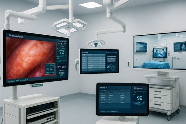
Selecting the right 4K surgical display1 goes beyond resolution. For minimally invasive surgery2, the features that truly impact clinical outcomes are those designed for the harsh reality of the operating room environment. This guide breaks down the five key pillars of a top-tier MIS display: visibility under surgical lights, signal reliability, true-to-life color fidelity, intuitive multi-image workflow, and practical hygiene and integration. Understanding these factors is essential for making an investment that enhances surgical precision and OR efficiency.
MIS-critical visibility: anti-glare optics, optical bonding, uniform luminance
Bright surgical lights often create debilitating glare on standard displays. This veiling glare washes out the image, obscuring critical anatomical details and compromising surgical safety.
Anti-glare protective glass combined with optical bonding is more important than on-paper contrast ratios. This pairing eliminates internal reflections and boosts perceived sharpness under intense OR lighting.
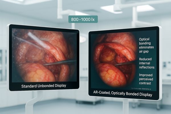
In the OR, with surgical lights often producing 800–1000 lux, a display’s "effective" contrast is what matters, not its theoretical spec. Veiling glare from ambient light can crush perceived contrast, making it difficult to distinguish between delicate tissue planes. The solution is twofold. First, anti-reflection (AR) coated protective glass3 minimizes surface reflections. Second, optical bonding—the process of laminating this glass directly to the LCD panel—eliminates the internal air gap. This removal of two reflective surfaces drastically reduces internal reflections and raises the Modulation Transfer Function (MTF)4, making fine details like vessel edges appear sharper and more defined. Surgeons perceive this as superior clarity, allowing them to work confidently without needing to crank the monitor’s brightness to overcome glare. The MS321PB is engineered with this philosophy, prioritizing real-world visibility to ensure the image remains crisp and clear during critical procedures.
True 4K@60 reliability in the OR: 12G-SDI, lockable connectors, low-latency paths
Signal loss during a procedure is a non-negotiable failure. Using consumer-grade cables like HDMI with extenders over long distances introduces multiple potential points of failure and signal degradation.
A single 12G-SDI cable provides a robust, low-latency 4K@60Hz signal path with locking connectors. This "fewer boxes, fewer surprises" approach is essential for mission-critical OR environments.
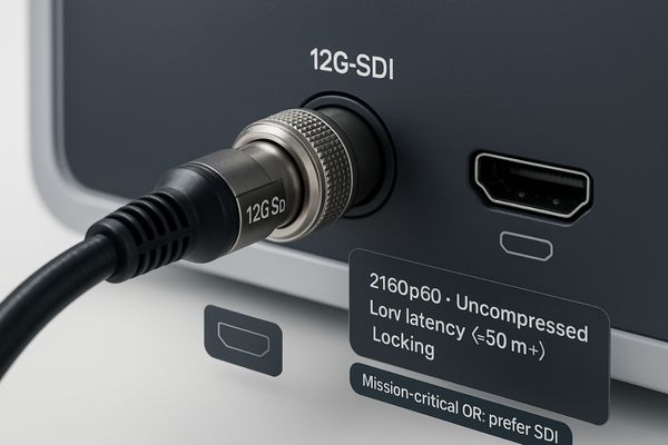
Delivering a stable, uncompressed 4K video5 signal at 60 frames per second is a core requirement for smooth surgical visualization. While HDMI is a common standard, it is not ideal for the OR. Its friction-based connectors can be accidentally dislodged, and running signals over 5 meters often requires extenders, which add complexity and latency. In contrast, 12G-SDI is purpose-built for professional video environments. A single 12G-SDI6 run carries uncompressed 2160p60 with lockable BNC and deterministic low latency over OR-grade coax; with qualified cabling, 50 m+ is common in practice. This streamlined signal path improves reliability by minimizing the number of components that could fail. In minimally invasive surgery, where every second counts, this built-in resilience is not a luxury; it is a fundamental safety feature.
Tissue-tone fidelity: BT.709/BT.2020 color, 10–12-bit pipeline, calibrated output
Inaccurate color reproduction can misrepresent tissue health. Displays that are overly saturated or exhibit color banding in subtle gradients can lead to clinical misinterpretation and reduce surgeon confidence.
Color in the OR is clinical, not cosmetic. True tissue-tone fidelity demands a calibrated output and a 10-bit or 12-bit pipeline to ensure smooth, accurate color gradients.
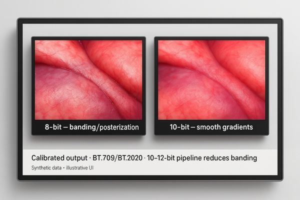
A surgeon’s ability to assess tissue perfusion, identify anatomical structures, and recognize abnormalities depends on accurate color reproduction. This is achieved through a combination of color space compliance7 and bit depth. A professional surgical display must be calibrated to standardized color gamuts, with BT.709 serving as the baseline for HD sources and BT.2020 as the target for UHD/4K content. More importantly, the display should have at least a 10-bit color processing pipeline8. This allows for over 1 billion colors, compared to the 16.7 million colors in an 8-bit system. The increased bit depth dramatically reduces color banding, resulting in smooth, natural gradients in low-contrast tissue folds and fluids. This level of fidelity ensures that what the surgeon sees on screen is a faithful representation of the surgical site, which is paramount for procedural safety and accuracy.
| Feature | 8-Bit Color | 10-Bit Color | Clinical Impact |
|---|---|---|---|
| Colors | 16.7 Million | 1.07 Billion | Reduces color banding in gradients |
| Application | Consumer Displays | Professional Surgical Displays | Improves tissue-tone fidelity |
| Result | Posterization, visible steps | Smooth, natural transitions | Higher diagnostic confidence |
Multi-image workflow for MIS: PIP/PBP/quad, quick OSD layouts per case
Fumbling with complex display menus during a procedure is disruptive and unsafe. Without an efficient way to manage multiple video sources, multi-image viewing features become a hindrance, not a help.
Multiview layouts only improve OR efficiency if each window can be independently configured and presets can be recalled instantly. Mapping layouts to specific procedures cuts down head-down time.
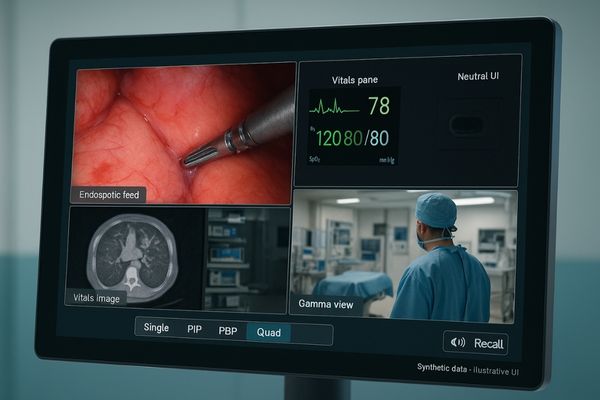
Modern MIS often requires viewing multiple sources simultaneously—such as the primary endoscopic camera, a secondary ultrasound feed, patient vitals, and radiological images. An effective surgical display must make this seamless. Features like Picture-in-Picture (PIP)9, Picture-by-Picture (PBP), and quad-view are essential, but their usability depends on the control interface. The best systems allow each window’s parameters (gamma, color, luminance) to be configured independently, ensuring each source is displayed accurately. Furthermore, the ability to save these layouts as one-touch presets10 in the on-screen display (OSD) or recall them via a footswitch is critical. By creating profiles for specific procedures like laparoscopy, arthroscopy, or ENT, the surgical team can switch views instantly without looking away from the operative field. This simple optimization prevents mode-switch errors and minimizes disruptions, directly contributing to a safer and more efficient workflow.
Hygiene & integration: flat, wipe-clean front, IEC 60601-1, arm-mount/VESA
Poorly designed hardware can slow down room turnover and create infection control risks. Cumbersome monitors with difficult-to-clean surfaces and non-standard mounts complicate OR setup and maintenance.
A flat, sealed front panel, IEC 60601-1 compliance, and standard VESA mounting are non-negotiable. These design features save time, improve safety, and deliver measurable ROI through faster room resets.
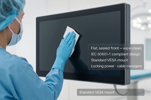
The physical design of a surgical display11 is as important as its electronic performance. A fully flat, sealed front surface with edge-to-edge protective glass is essential for effective and rapid cleaning between procedures, supporting strict infection control protocols. Compliance with the IEC 60601-112 medical safety standard ensures the device is safe for use in the patient vicinity. From an integration standpoint, a standard VESA 100×100 mounting pattern provides compatibility with a wide range of surgical booms and wall mounts, simplifying installation. Thoughtful design elements like integrated cable routing and locking power connectors prevent clutter and reduce the risk of inadvertent disconnections. These "invisible" features, found on displays like the MS322PB, accumulate into significant time savings during room turnover. In a busy surgical department, faster resets and more consistent setups lead directly to higher throughput and a tangible return on investment.
Conclusion
Choose a 4K surgical display that excels in real-world visibility, signal reliability, color fidelity, workflow efficiency, and hygienic design to truly enhance surgical outcomes. ✨
👉 For expert guidance and tailored Reshin surgical display recommendations, contact martin@reshinmonitors.com.
-
Explore how a 4K surgical display can enhance surgical precision and improve outcomes in minimally invasive surgeries. ↩
-
Discover the latest techniques and technologies in minimally invasive surgery that can improve patient outcomes and surgical efficiency. ↩
-
Explore how AR coatings enhance visibility and reduce glare in surgical environments, ensuring precision during operations. ↩
-
Learn about MTF’s role in image clarity and detail, crucial for surgeons needing accurate visual information. ↩
-
Learn about the significance of uncompressed 4K video in enhancing surgical procedures and patient safety. ↩
-
Explore the benefits of 12G-SDI to understand why it’s preferred for high-quality video in professional settings. ↩
-
Understanding color space compliance is crucial for ensuring accurate color reproduction in surgical displays, enhancing surgical precision. ↩
-
Exploring the significance of a 10-bit color processing pipeline reveals how it improves color fidelity and surgical outcomes. ↩
-
Explore how PIP enhances surgical displays for better multitasking and efficiency in the operating room. ↩
-
Learn how one-touch presets streamline operations, allowing for quick adjustments and improved focus during procedures. ↩
-
Explore this link to understand the essential features and benefits of surgical displays in medical settings. ↩
-
Learn about the IEC 60601-1 standard to ensure safety and compliance in medical devices, crucial for patient care. ↩