Analyzing complex hematology images on a standard computer monitor is risky. Inaccurate color and poor detail can easily mask the subtle cellular abnormalities that are critical for a correct diagnosis.
In hematology, accurate visualization of blood smears, bone marrow biopsies, and flow cytometry plots relies on medical-grade displays. With calibrated resolution, precise color fidelity, and luminance stability, they reveal subtle variations and cellular details. By combining DICOM GSDF grayscale standards with ICC-based color management, these displays ensure diagnostic consistency, improve workflow efficiency, and reduce eye strain during long hours of analysis.
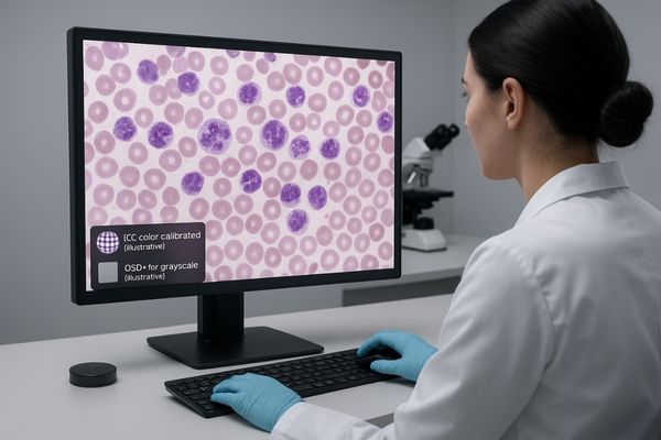
The field of hematology is profoundly visual. From identifying blast cells in a bone marrow smear to gating populations in a flow cytometry plot, every decision depends on the accurate interpretation of visual data. As laboratories increasingly adopt digital workflows, the display monitor has become as critical as the microscope itself. It serves as the final window through which all digitally captured information is viewed, analyzed, and interpreted. Using a consumer-grade screen in this context introduces an unacceptable level of variability and risk. This article will delve into the specific requirements of hematology imaging1, explaining why medical-grade displays2 with their calibrated performance, advanced features, and ergonomic design are not just a preference but a clinical necessity for modern laboratory practice.
Hematology Imaging Tasks and Visual Demands
Not all hematology images are the same. A peripheral blood smear and a whole-slide biopsy image present completely different visual challenges, requiring different display capabilities for accurate interpretation.
Hematology imaging includes blood smears needing precise color fidelity, whole-slide images demanding high resolution for zooming, and flow cytometry plots. Digital morphology analyzers now improve efficiency and standardize results.
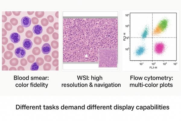
The visual demands in hematology are highly varied, dictated by the specific diagnostic task. Peripheral blood smears and bone marrow aspirates stained with chromogenic dyes like Wright–Giemsa require exceptional color fidelity to differentiate between cell types and identify subtle abnormalities in nuclear chromatin or cytoplasmic granules. The display must render these colors consistently to avoid misinterpretation. In contrast, digital pathology3 using whole-slide imaging (WSI) of bone marrow biopsies introduces the challenge of scale. Pathologists need to navigate vast digital landscapes, seamlessly zooming from a low-power overview to high-power magnification to inspect individual cells. This requires a high pixel count4 to maintain image sharpness and prevent the loss of micro-features during magnification. Furthermore, modern digital morphology analyzers, which automate the analysis of blood smears, have demonstrated significant real-world gains in both efficiency and precision. By standardizing results and reducing manual labor, they depend on high-quality displays to present their findings for review and verification, making the display a core component of this increasingly automated workflow.
Display Performance Requirements: Resolution, Color, Contrast, Stability
Using a screen with poor resolution or unstable brightness can obscure critical cellular details. This variability can lead to diagnostic uncertainty and potential errors in cell classification over time.
Displays for hematology need at least Full HD resolution, with 3-8MP preferred for primary reading. High contrast, stable luminance, and hardware calibration are essential for revealing subtle chromatic differences consistently.
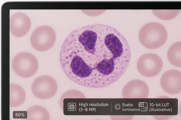
To meet the demanding needs of hematology, a medical-grade display5 must possess specific performance characteristics that far exceed those of consumer monitors. Resolution is fundamental. While a Full-HD (2MP) display may be adequate for basic review, primary diagnostic reading and whole-slide navigation benefit significantly from higher pixel densities of 3MP to 8MP. This ensures that the fine details of cellular morphology—such as nuclear shape, chromatin pattern, and cytoplasmic granules—remain crisp and well-defined even at typical working distances. High contrast and stable luminance are equally critical for revealing subtle chromatic variations between different stains and textures within the cytoplasm. To achieve this, medical displays utilize hardware-stabilized LED backlights that maintain consistent brightness and color temperature over thousands of hours of use. This long-term stability is locked in through regular calibration. Hardware calibration, which leverages internal 10-bit or higher lookup tables (LUTs), offers far greater accuracy than software-only solutions by minimizing tonal distortion and ensuring that the display’s full color gamut is rendered with precision.
Standards: DICOM GSDF for Grayscale and ICC-Based Color Management
Without established standards, the same image would look different on every monitor. This inconsistency undermines the reliability of remote consultations, longitudinal studies, and inter-reader agreement.
Grayscale content is calibrated to the DICOM Part 14 standard for perceptual uniformity. Color images utilize ICC-based management for device-independent appearance, allowing one workstation to render all content consistently.
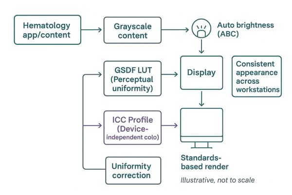
To ensure reliable and repeatable image interpretation, medical displays adhere to internationally recognized standards for both grayscale and color rendering. Although hematology is a color-rich discipline, some imaging data may have grayscale components or be viewed using pseudo-color, making grayscale standards relevant. Any grayscale content should be calibrated to the DICOM (Digital Imaging and Communications in Medicine)6 Part 14 Grayscale Standard Display Function (GSDF). This standard is based on the Barten model of human visual perception and ensures that each step in the grayscale is perceptually uniform, making subtle differences in tone equally noticeable. For the vast majority of hematology tasks, color management is paramount. This is achieved using ICC (International Color Consortium) profiles7. Standards like sRGB or Adobe RGB define a device-independent color space, and the display’s calibration software creates a specific ICC profile for the monitor. Regulatory discussions explicitly describe the process of transforming sRGB content to the display’s unique profile during calibration. By embracing both DICOM GSDF for grayscale and ICC workflows for color, a single hematology workstation can render all types of content with proven consistency.
Uniformity, Anti-Reflection, and Ambient-Light Control
Glare from room lights and uneven brightness across the screen can mask subtle details. This forces users into awkward positions or constant image adjustments, causing fatigue and potential diagnostic errors.
Ambient lighting affects perceived contrast, so medical displays use advanced anti-reflection optics and ambient light sensors to preserve image quality. Uniform luminance across the panel is essential for consistent scanning.
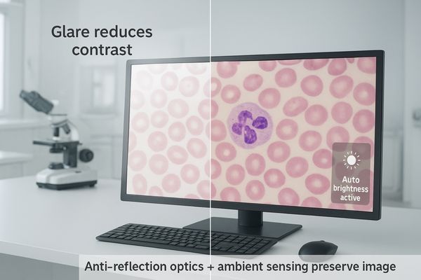
The environment in which a display is viewed has a significant impact on perceived image quality. Ambient room lighting can wash out contrast and create distracting reflections on the screen surface. For this reason, laboratory and reading room guidance often recommends controlled ambient light levels of around 25–40 lux. Medical grade displays are designed to perform optimally within this context. They incorporate advanced anti-reflection and anti-glare optical coatings that dramatically reduce specular reflections from overhead lights and windows, thereby preserving image contrast and color saturation. Many models also include a built-in ambient light sensor8 that can automatically adjust the display’s brightness to compensate for changes in room lighting, ensuring a consistent viewing experience. Another critical feature is luminance uniformity. Specialized internal hardware, often called a Digital Uniformity Equalizer (DUE)9, ensures that brightness is consistent across the entire panel, from the center to the corners. This is vital when visually scanning a large digital smear or navigating a whole-slide image, as it guarantees that the appearance of cells is consistent regardless of their position on the screen.
Workflow & Ergonomics for Long-Duration Use
Spending hours analyzing digital slides on a poorly designed workstation leads to significant physical strain. This fatigue not only impacts well-being but can also degrade diagnostic performance over time.
Hematology analysts face high rates of digital eye strain. Ergonomic features like flicker-free backlights, calibrated luminance, and fully adjustable stands are essential for reducing fatigue during long work sessions.
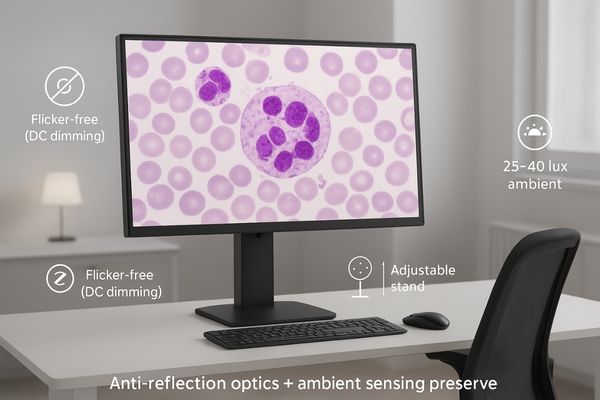
The work of a hematology analyst or pathologist involves long periods of intense visual concentration. This places them at high risk for digital eye strain10 and musculoskeletal discomfort, issues that studies show are highly prevalent among digital imaging professionals. Medical-grade displays are engineered with specific ergonomic features to mitigate these risks. One of the most important is a flicker-free backlight. Unlike consumer monitors that often use Pulse-Width Modulation (PWM) for dimming, which can cause imperceptible but fatiguing flicker, medical displays use DC dimming to provide a stable, comfortable light source. Properly calibrated luminance is also key, as it prevents the screen from being overly bright or dark relative to the ambient environment, a common cause of eye strain. Finally, the physical stand is a critical ergonomic component. A fully adjustable stand that allows for height, tilt, swivel, and pivot adjustments enables users to position the screen perfectly for their posture, reducing neck and back strain. Together, these features create a more comfortable and sustainable work environment, helping to maintain high levels of accuracy and concentration throughout the day.
Hematology Workstation Integration: Microscope-Camera and Multi-Monitor
Integrating live feeds and multiple monitors can lead to inconsistent color and distracting lag. This makes collaboration, teaching, and efficient multitasking difficult and unreliable.
Workstation integration demands color-accurate displays for live microscope feeds and seamless multi-monitor setups. This enables reliable remote collaboration, tele-microscopy, and efficient analysis of multi-modal data.
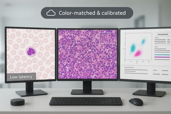
A modern hematology workstation is often a complex setup, integrating live microscope camera feeds alongside static images and patient data. In these environments, display performance is key to a seamless workflow. For live microscope viewing, whether for teaching, consultation, or tumor boards, a large-format, color-accurate display is essential. A display like the MS551P with its low lag and precise color rendering ensures that multiple observers, whether local or remote, are seeing an identical and faithful representation of what is under the lens. Studies on tele-microscopy have shown that this capability improves turnaround times by allowing remote experts to review fields of view with confidence. The same principle applies to multi-monitor setups, where an analyst might compare a current slide to a prior one. The displays must be perfectly calibrated and matched so that color and brightness are consistent across screens. In flow cytometry, a large, high-resolution monitor like the MS431P can be vital for accurately rendering complex multi-color plots, allowing for precise adjustment of compensation and gating, which is fundamental to achieving an accurate analysis.
Clinical Impact and QA: Accuracy, TAT, and Routine Calibration
Without consistent quality assurance, even the best display will degrade over time. This slow decay in performance can introduce subtle errors into a lab’s diagnostic process without anyone noticing.
Display quality directly impacts diagnostic accuracy and turnaround time. Routine quality assurance, including luminance testing and DICOM conformance checks, is required to maintain this performance and ensure reliable results.
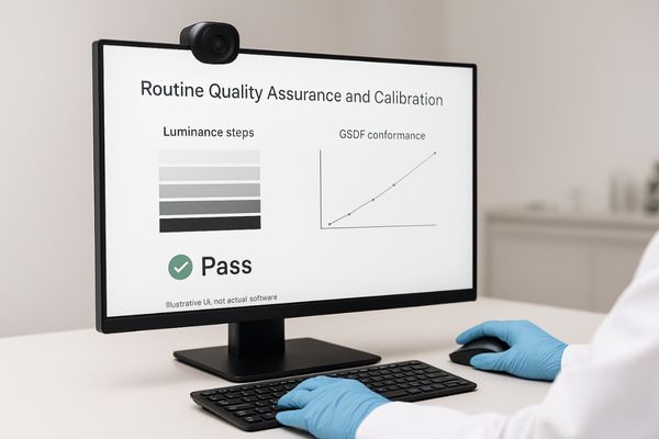
The clinical impact of a high-quality display, maintained through a rigorous quality assurance (QA) program11, is significant. Optimized hematology workstations that combine a high-resolution, color-calibrated display with a controlled ambient light environment can demonstrably improve the recognition of subtle chromatic and textural cues in cells. This leads to faster, more consistent morphology calls and strengthens inter-reader agreement, ultimately improving diagnostic accuracy and patient safety. However, this performance can only be sustained through routine QA. Professional practice parameters require, at minimum, annual testing of the display’s luminance characteristics—including its performance with ambient light contributions—and verification that it maintains conformance with the DICOM GSDF standard12. This routine calibration is not a "set it and forget it" task. It is an ongoing process of verification that ensures the display continues to perform as a precision medical instrument. This commitment to quality assurance is what underpins diagnostic confidence and ensures that the laboratory’s results are both accurate and defensible over the long term.
Conclusion
Medical-grade displays are essential tools in digital hematology, providing the calibrated color, resolution, and stability required for confident and consistent analysis of cellular morphology.
-
Understanding the requirements for hematology imaging is crucial for ensuring accurate diagnostics and effective laboratory practices. ↩
-
Exploring the importance of medical-grade displays can help you appreciate their role in enhancing diagnostic accuracy and patient safety. ↩
-
Explore this link to understand how digital pathology enhances diagnostic processes and improves efficiency in hematology. ↩
-
Learn why high pixel count is crucial for maintaining image quality and detail in pathology, ensuring accurate diagnoses. ↩
-
Explore this link to understand the essential features of medical-grade displays that enhance diagnostic accuracy in hematology. ↩
-
Explore this link to understand DICOM’s role in standardizing medical imaging, ensuring accurate grayscale and color rendering. ↩
-
Learn about ICC profiles to see how they enhance color management in medical displays, crucial for accurate image interpretation. ↩
-
Understanding ambient light sensors can enhance your knowledge of display technology and improve your viewing experience. ↩
-
Exploring DUE technology will help you appreciate how displays maintain consistent brightness and image quality. ↩
-
Understanding digital eye strain can help you implement effective strategies to reduce discomfort and improve your work experience. ↩
-
Understanding the role of QA programs can enhance your knowledge of maintaining high standards in clinical diagnostics. ↩
-
Exploring the DICOM GSDF standard will provide insights into display calibration and its impact on medical imaging accuracy. ↩