Choosing the wrong display for a clinical department can lead to diagnostic errors or surgical complications. A monitor optimized for one environment will fail in another, creating unacceptable risks.
This article details the critical differences in selecting displays for radiology versus the operating room. We will compare requirements for resolution, brightness, color accuracy, durability, connectivity, and ergonomics to guide informed purchasing decisions and ensure optimal clinical performance.
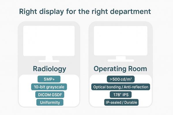
The selection of a medical display1 is a decision with direct clinical consequences. While both radiologists and surgeons rely on visual information, the nature of their tasks dictates vastly different technological priorities. The radiologist’s world is one of detailed analysis, demanding absolute precision in grayscale and color to interpret static images. They require tools optimized for diagnostic confidence. In contrast, the surgeon’s environment is dynamic and collaborative, demanding real-time video performance, high brightness to overcome harsh lighting, and ruggedness to survive a sterile environment. Understanding this fundamental distinction is crucial. For instance, a general-purpose diagnostic monitor2 like the MD33G is engineered for the controlled lighting of a reading room, prioritizing pixel-perfect clarity and DICOM compliance above all else. Attempting to use this same display in a brightly lit OR would compromise visibility, just as using a surgical display for primary diagnosis would sacrifice the nuanced detail required for accurate interpretation.
Radiology displays demand high resolution and grayscale depth
A radiologist can miss a subtle hairline fracture or a small nodule on a low-resolution display. This diagnostic ambiguity is unacceptable and can delay critical patient treatment.
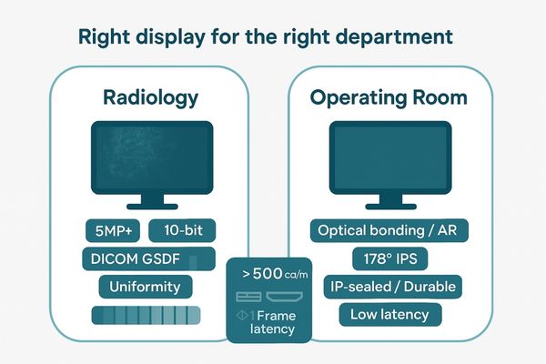
For diagnostic confidence, radiology workstations require ultra-high resolution, often 5MP or more, combined with 10-bit grayscale depth. This is essential to visualize faint pathologies and ensure consistent image interpretation according to the DICOM standard.
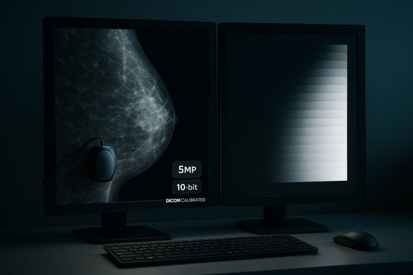
In radiology, the display’s resolution is directly linked to its diagnostic power. The goal is to see the smallest possible detail with clarity. This is why specific modalities have minimum resolution standards. For mammography, where detecting microcalcifications is key, a 5MP grayscale monitor3 like the MD52G is essential. Its high pixel density ensures that these tiny, critical indicators are not lost due to pixel averaging on a lower-resolution screen. This is complemented by 10-bit grayscale, which provides 1,024 distinct shades of gray compared to the 256 of a standard 8-bit display. This expanded tonal range allows for the subtle differentiation of soft tissues, which is crucial for identifying tumors or other abnormalities. Adherence to the DICOM Part 14 standard4 ensures these grayscale values are rendered consistently across different displays and viewers, forming a reliable foundation for clinical decisions.
| Modality | Minimum Recommended Resolution | Primary Rationale |
|---|---|---|
| Mammography | 5MP (Grayscale) | Visualization of microcalcifications |
| CT/MRI (Chest) | 3MP | Detection of small nodules and fine structures |
| PET/CT Fusion | 3MP – 5MP (Color) | Combining anatomical and metabolic detail |
| General X-Ray | 2MP | Basic review of skeletal and chest radiography |
Operating room displays require high brightness and wide viewing angles
A standard display becomes washed out and unreadable under intense surgical lamps. This forces the surgical team to squint or reposition, compromising focus and collaboration during a procedure.
In the OR, high brightness (often over 500 cd/m²) and advanced anti-reflective finishes are mandatory for maintaining image clarity. Wide viewing angles from an IPS panel ensure visual consistency for the entire surgical team.
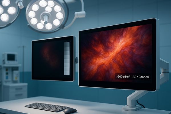
The operating room is a uniquely challenging visual environment. The powerful, focused lamps used to illuminate the surgical site create intense ambient light that can overwhelm a conventional display. To counteract this, surgical monitors must deliver high luminance5. A monitor like the MS321PB is engineered with a high-efficiency backlight system to produce brightness levels that cut through the glare, providing a clear, high-contrast image. This is often enhanced by optical bonding, which eliminates the air gap between the panel and protective glass to reduce internal reflections and improve perceived contrast. Equally important are the viewing angles. The surgical team is distributed around the patient, and each member must see the same accurate image. IPS (In-Plane Switching) panel technology6 is the standard here, providing 178-degree viewing angles without the color or contrast shift common on lesser panels. This ensures the surgeon, assistants, and nurses all share a single, consistent visual reference point.
Color accuracy is critical for radiology diagnostic confidence
Inaccurate color on a display can obscure or mimic pathology in modalities like PET/CT. A radiologist might misinterpret the metabolic activity of a tumor if the monitor’s color reproduction is flawed.
Advanced calibration systems are vital for radiology displays to maintain luminance stability and color uniformity over time. This ensures that diagnoses are based on true image data, not on monitor-induced artifacts or color shifts.
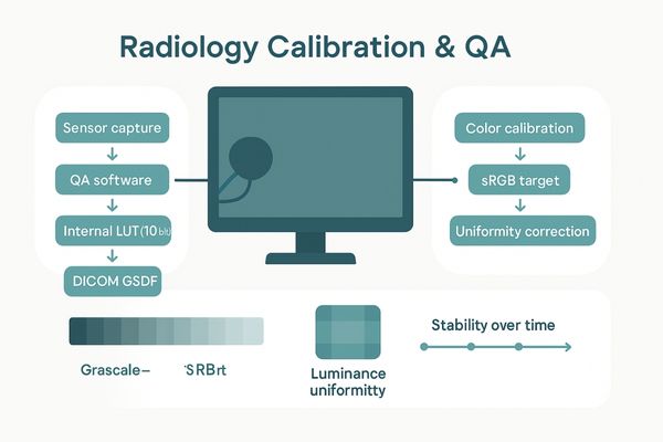
While grayscale is foundational, accurate color reproduction7 has become indispensable with the rise of multi-modality imaging. In PET/CT fusion, for example, color maps are used to represent the metabolic uptake of a radiotracer. The precise hue and saturation of the color provide quantitative information about tissue activity. A display that cannot render these colors accurately compromises the entire diagnostic premise. To prevent this, advanced diagnostic monitors8 like the MD26C incorporate sophisticated calibration technologies. This includes a built-in front sensor that periodically measures the screen’s output and an accompanying software suite that automatically adjusts the display to maintain perfect alignment with standards like DICOM and sRGB. Furthermore, uniformity correction technology ensures that color and luminance are identical from the center of the screen to the edges. This constant, automated quality assurance gives radiologists the confidence that the image they are seeing is a faithful representation of the patient data.
Surgical monitors must withstand rigorous cleaning protocols
The operating room demands absolute sterility. A surgical monitor must be cleaned frequently with harsh chemical disinfectants whose vapors and liquids can damage consumer-grade plastics and seep into unsealed enclosures.
Durability is a top priority for OR monitors. They must have IP-rated, sealed enclosures and chemically resistant coatings that can endure frequent and aggressive disinfection without degrading, ensuring both patient safety and long-term equipment reliability.
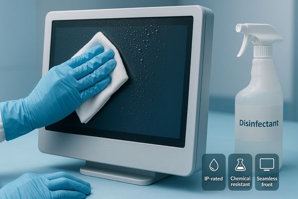
A https://reshinmonitors.com/top-medical-display-manufacturers-2025/9 is, first and foremost, a piece of medical equipment that lives in a sterile field. This reality dictates its physical design. The entire housing must be built to withstand repeated wipe-downs with strong cleaning agents. Consumer-grade plastic enclosures would quickly become brittle and crack under such a regimen. Therefore, monitors like the MS270P feature rugged metal or medical-grade polymer housings and seamless, bezel-free front designs. The front panel is a single piece of protective glass that eliminates seams or gaps where biological contaminants could accumulate. Furthermore, these monitors carry an Ingress Protection (IP) rating10, which certifies their level of resistance to the intrusion of liquids and dust. A sealed chassis and protected I/O ports prevent fluids from damaging the internal electronics, a common point of failure for non-medical displays used in the OR. This robust, sterile-friendly construction is non-negotiable for infection control and long-term reliability.
Connectivity needs differ between diagnostic and surgical workflows
A radiologist needs stable, high-bandwidth connections to a PACS workstation. A surgeon needs versatile, low-latency inputs from multiple live video sources like an endoscope, ultrasound, and OR camera.
Surgical monitors require a wide range of inputs and the ability to switch between them rapidly. This supports the dynamic integration of endoscopy, ultrasound, navigation systems, and other live imaging sources during a procedure.
While both environments require reliable signal connections, their workflows demand different input configurations. A radiology reading room typically uses a stable, high-bandwidth DisplayPort connection11 from a dedicated PACS workstation. The setup is static. In contrast, the modern operating room is a hub of live video feeds. A single large-format surgical display like the MS550P must be able to accept simultaneous inputs from a 4K endoscopic camera12 via 12G-SDI, a mobile C-arm via HDMI, and a room camera via another input. The monitor’s internal processing must handle these various signals with near-zero latency to maintain the surgeon’s hand-eye coordination. The ability to quickly switch between these sources or display them simultaneously using Picture-in-Picture (PIP) or Picture-by-Picture (PBP) layouts is a critical feature. This input versatility and rapid processing are essential for the fluid, multi-source nature of contemporary surgical procedures.
| Feature | Radiology Display Priority | Operating Room Display Priority |
|---|---|---|
| Inputs | DisplayPort | SDI (3G/12G), HDMI, DP, DVI |
| Latency | Not critical for static images | Very Low (<1 frame) is essential |
| Source Mgmt | Single-source from PACS | Multi-source switching (PIP/PBP) |
Ergonomic design impacts efficiency in both environments
Poor ergonomics can lead to repetitive strain injuries for radiologists who spend hours reading studies. In the OR, a non-adjustable monitor can impede visibility for parts of the surgical team.
Ergonomics are crucial in both settings, but for different reasons. Radiologists need highly adjustable stands to minimize strain, while OR monitors require flexible boom mounting to ensure optimal positioning for a collaborative team.
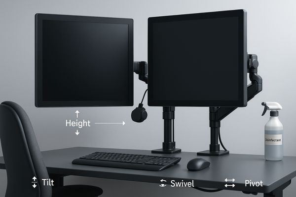
Ergonomic design13 directly contributes to clinical performance by reducing fatigue and improving comfort. For a radiologist, who spends most of their day analyzing images, this means having a highly adjustable monitor stand. The ability to easily control height, tilt, swivel, and pivot allows the user to maintain a neutral posture, reducing the risk of neck and back strain. Specialized multi-head displays, such as the dual-screen MD45C, are engineered to present a seamless desktop and minimize bezel gap, allowing for efficient comparison of prior and current studies. In the operating room, ergonomics are about positioning and accessibility. Surgical displays are rarely placed on a desk; instead, they are mounted on ceiling booms or mobile carts. VESA mount compatibility14 is essential, as is a lightweight yet durable construction that allows the boom to be moved and positioned easily without drift. This flexibility ensures the screen can be placed in the ideal line of sight for the entire team.
Reshin tailors display specifications to department-specific needs
Using a single display model across a hospital leads to clinical compromises. A jack-of-all-trades display is a master of none, failing to meet the specific demands of either radiology or surgery.
Our product lines for radiology and the OR are purpose-built to address these contrasting requirements. This ensures full compliance with relevant standards like DICOM and surgical safety protocols, providing optimized tools for each unique clinical task.
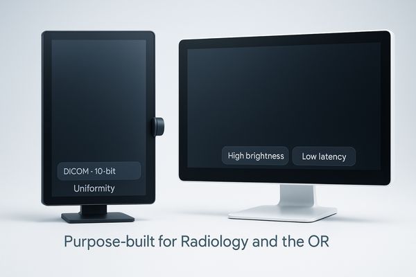
My philosophy is grounded in creating purpose-built tools. We do not attempt to create a single monitor to serve all departments. Instead, we engineer distinct product families that address the specific challenges of each clinical environment. Our diagnostic monitors15 are designed for the controlled conditions of the reading room, focusing on impeccable image fidelity. We develop unique solutions like the 34-inch panoramic MD51CHY, which provides a 5MP-equivalent resolution in an ultrawide format to streamline workflow for chest radiology and other specialties. Conversely, our https://reshinmonitors.com/top-medical-display-manufacturers-2025/16 are built for the demanding environment of the OR. They feature high-brightness panels, sealed metal enclosures, and the low-latency processing required for live video. By focusing on these department-specific needs, we provide clinicians with tools that are not just compliant, but are truly optimized for their work, enhancing efficiency, safety, and diagnostic confidence.
Conclusion
Selecting the right medical display requires matching its specific features—resolution, brightness, and durability—to the distinct demands of either the radiology reading room or the operating room.
📧 Need help choosing displays tailored to radiology or surgical applications? Contact Martin at martin@reshinmonitors.com for expert guidance from Reshin.
- Explore this link to understand the critical factors in choosing the right medical display for clinical settings. ↩
- This resource will provide insights into essential features that enhance diagnostic accuracy for radiologists. ↩
- Explore this link to understand how a 5MP grayscale monitor enhances diagnostic accuracy in radiology. ↩
- Learn about the DICOM Part 14 standard to see how it ensures consistency in medical imaging across different devices. ↩
- Understanding high luminance is crucial for ensuring optimal visibility in surgical environments, enhancing patient safety. ↩
- Exploring IPS technology will reveal its advantages in providing consistent image quality, essential for surgical teams. ↩
- Understanding color reproduction is crucial for accurate diagnostics in medical imaging, ensuring reliable patient assessments. ↩
- Exploring advanced diagnostic monitors reveals how technology enhances image quality and diagnostic accuracy in healthcare. ↩
- Understanding the features of surgical monitors can help ensure the right choice for medical environments. ↩
- Exploring the significance of IP ratings can enhance your knowledge of equipment durability and safety. ↩
- Understanding DisplayPort connections can enhance your knowledge of reliable signal transmission in medical settings. ↩
- Exploring the advantages of 4K endoscopic cameras can provide insights into advancements in surgical technology. ↩
- Explore how ergonomic design enhances clinical performance by reducing fatigue and improving comfort for healthcare professionals. ↩
- Learn about VESA mount compatibility and its significance in ensuring optimal positioning and accessibility of surgical displays. ↩
- Explore this link to discover top-rated diagnostic monitors tailored for various clinical settings, enhancing image fidelity and workflow. ↩
- Check out this resource to learn about essential features of surgical displays that ensure optimal performance in demanding operating room conditions. ↩


