Noisy, distorted images can obstruct critical diagnostic information. This interference compromises clinical decisions. How do medical displays ensure the pristine clarity essential for healthcare?
Medical displays achieve signal noise reduction through a combination of robust hardware design, advanced signal processing algorithms, effective shielding and grounding techniques, and the use of high-quality, specialized components that minimize interference and enhance image clarity.
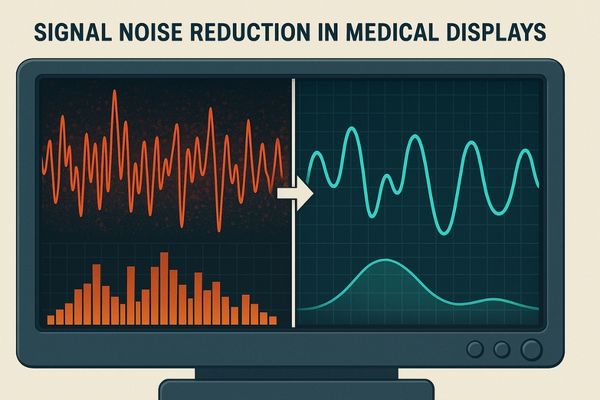
The clarity of a medical image can be the difference between an accurate diagnosis and a missed opportunity. In the complex electromagnetic environment of a modern hospital, ensuring a clean signal1 is paramount. This article will delve into the specific methods and technologies medical displays2 employ to combat noise and deliver pristine images3. We will explore how these systems work to provide reliable visual information to medical professionals.
What types of noise commonly affect medical display signals?
Medical images can sometimes flicker or show unexpected artifacts. These issues can obscure crucial details. It is important to understand what insidious noise sources corrupt medical display signals in demanding clinical environments.
Medical display signals are commonly affected by electromagnetic interference (EMI) and radio-frequency interference (RFI) from nearby equipment. [Signal](https://reshinmonitors.com/surgical-monitor-no-signal-troubleshooting/ “Signal”) jitter causing instability, and signal degradation or loss during transmission also degrade image quality, particularly in busy clinical environments.
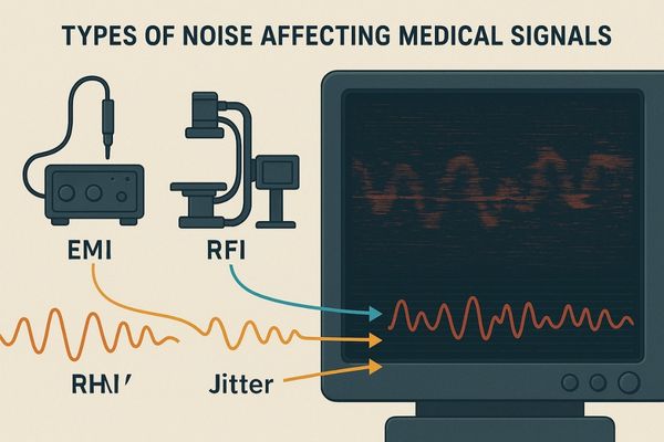
Several types of noise can degrade the quality of signals reaching a medical display. Electromagnetic Interference (EMI)4 is a common culprit. It originates from electrical and electronic devices that radiate electromagnetic fields. Sources include electrosurgical units, motors, and even fluorescent lighting. EMI can manifest as visible patterns, lines, or distortions on the screen. Radio-Frequency Interference (RFI)5 is similar but specifically refers to interference from radio waves. Wireless communication devices, RFID systems, and other medical equipment broadcasting radio frequencies can introduce RFI. This may appear as speckles, wavy patterns, or “snow” on the image. Signal jitter is another problem. This refers to undesirable variations in the timing of digital signals. Poor quality cables, connectors, or internal clocking mechanisms in the source or display can cause jitter. It results in an unstable or shaky image, and in severe cases, loss of synchronization. Transmission loss, or signal attenuation, occurs as signals weaken over distance or pass through multiple connections. Long cable runs or poor-quality connectors can lead to a dim image, color shifts, or a complete loss of signal. Operating rooms and intensive care units are particularly prone to these interferences due to the high concentration of active electronic systems.
How do hardware designs contribute to signal noise suppression?
The internal components of a display play a significant role. Frustration can arise from subpar image quality. One might wonder if the display’s hardware is the source of the problem. Discovering specific design choices can reveal how signal noise is effectively silenced.
Hardware designs in medical displays contribute to noise suppression by incorporating low-noise power supplies and precision digital clocks for stable timing. They also use differential signal transmission to reject common-mode noise and internal signal equalizers to boost signal integrity.
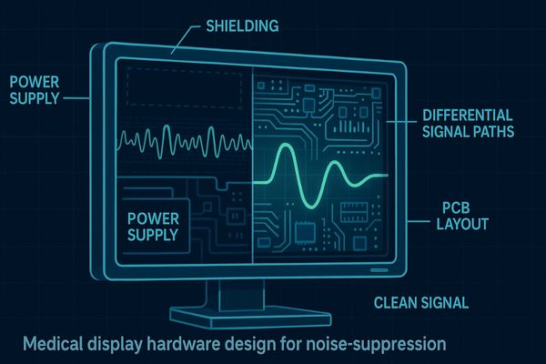
The hardware architecture of a medical display is fundamental to its ability to resist and suppress noise. Manufacturers of premium medical displays incorporate several key design strategies. Low-noise power supplies6 are critical. These units are engineered to provide clean, stable Direct Current (DC) power to the display’s sensitive electronics. They filter out noise and fluctuations from the AC mains supply, preventing power-line interference from corrupting video signals. Precision digital clocks are also essential. These provide highly accurate timing signals for synchronizing data transmission and pixel rendering. Stable clocking minimizes signal jitter, ensuring a crisp and stable image. Differential signal transmission7 is a widely used technique. Standards like LVDS (Low-Voltage Differential Signaling) and TMDS (Transition Minimized Differential Signaling), used in HDMI and DVI, transmit signals using pairs of wires carrying equal and opposite signals. The receiver subtracts one signal from the other. This process effectively cancels out common-mode noise, which is interference picked up equally by both wires. Furthermore, internal signal equalizers8, also known as re-drivers or re-timers, are often built into the display’s input circuitry. These components actively reshape and amplify weakened or distorted signals. This is particularly important for signals traveling over long cables, compensating for transmission loss and some forms of high-frequency jitter. The quality of passive components, such as capacitors and inductors, and meticulous Printed Circuit Board (PCB) layout also contribute significantly to minimizing internal noise generation and susceptibility.
What role do signal processing algorithms play in image noise reduction?
Even with robust hardware, some noise can persist. Grainy images might obscure crucial details. Unexpected color spots can mar the view. It is useful to uncover how smart algorithms clean up medical visuals effectively.
Signal processing algorithms in medical displays reduce image noise by identifying and filtering out non-structural artifacts like speckles and random color spots. They use spatial and temporal filtering techniques to enhance image clarity and uniformity without sacrificing diagnostic detail.
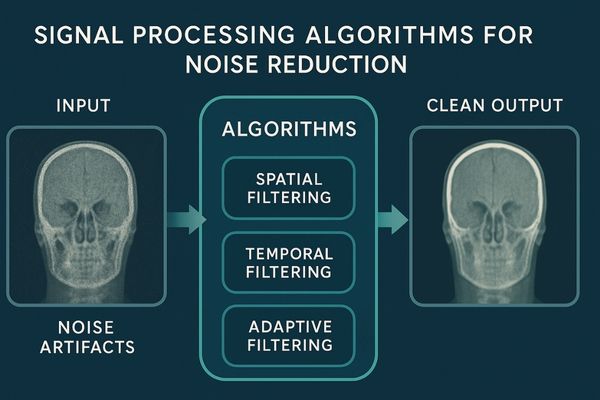
Advanced signal processing algorithms are a vital tool in the fight against image noise9 in medical displays. These sophisticated software routines, often implemented in dedicated hardware chips, analyze the incoming video signal to detect and mitigate various types of non-structural noise. This noise can include speckle noise, which appears as a granular pattern, salt-and-pepper noise characterized by random bright or dark pixels, and random color noise or spots. Spatial filtering is one common approach. These filters operate on a single image frame, examining the values of neighboring pixels to identify and smooth out noise. For example, a median filter replaces each pixel’s value with the median value of its surrounding pixels, which is effective against impulse noise like salt-and-pepper. Gaussian filters apply a weighted average, smoothing noise while trying to preserve edges. Temporal filtering, on the other hand, utilizes information from multiple frames. Techniques like frame averaging or more advanced motion-compensated temporal filtering can be very effective at reducing random noise, especially in static or slowly moving parts of an image. Adaptive filtering techniques are also employed; these algorithms adjust their filtering strength and type based on the local characteristics of the image, applying stronger filtering in noisy, uniform areas and lighter filtering near edges and details to preserve diagnostic information. The key challenge and goal of these algorithms is to reduce perceptible noise and enhance clarity without blurring or removing fine, diagnostically relevant details.
Table 1: Common Image Noise Filtering Techniques
| Filter Type | Primary Target Noise | Basic Principle | Potential Side Effect |
|---|---|---|---|
| Median Filter | Salt-and-pepper, impulse noise | Replaces pixel with median of neighbors | Some edge/detail smoothing |
| Gaussian Filter | General random noise | Weighted average of neighboring pixels | Blurring if too strong |
| Bilateral Filter | General random noise | Weighted average, considering spatial distance and intensity | Preserves edges better |
| Temporal Averaging | Random noise (static scenes) | Averages pixel values across multiple frames | Motion blur if not adapted |
| Anisotropic Diffusion | General noise, edge preserving | Smooths within regions, not across strong edges | Computationally intensive |
These algorithms are carefully tuned to balance noise reduction with the preservation of essential image content.
How does shielding and grounding affect display signal quality?
External interference can wreck a display’s clarity. A setup might be vulnerable to disturbances from other equipment. Understanding how physical protection helps maintain pure signals is important for reliable performance.
Proper shielding, using materials like metal enclosures, and effective grounding, which isolates signal paths from power grounds, are crucial. They protect display signals from external electromagnetic interference (EMI), ensuring high-fidelity image transmission and integrity.
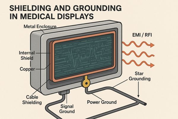
Shielding and grounding are fundamental physical measures for protecting display signals from external interference. Shielding10 involves enclosing sensitive electronic components and signal pathways within materials that block or attenuate electromagnetic fields. Typically, conductive materials like aluminum, steel, or specialized alloys such as mu-metal (for magnetic shielding) are used. This can take the form of a complete metal enclosure for the display, internal shielding around specific circuits (like the video processing board or power supply), and shielded cables. High-quality video cables, for instance, often feature braided copper shields and foil wraps around the signal conductors to prevent external EMI and RFI from corrupting the signal. The principle is similar to that of a Faraday cage11, which blocks external static and non-static electric fields. Grounding12 is equally important. It provides a common, stable electrical reference point (ground potential) for all parts of the system and a safe path for unwanted electrical currents to dissipate. In medical displays, careful grounding schemes are implemented. This includes separating sensitive signal grounds from potentially noisy power supply grounds to prevent noise from coupling between them. Star grounding, where all grounds connect to a single central point, can help avoid ground loops, which are a common source of hum and interference. The design of connectors and their connection to the chassis and cable shields also plays a vital role in maintaining ground integrity and effective shielding across interfaces. Together, robust shielding and meticulous grounding form a critical defense against the pervasive electromagnetic noise found in clinical settings.
Table 2: Shielding Materials and Their Applications
| Material | Shielding Property | Common Application in Displays |
|---|---|---|
| Aluminum | Good EMI/RFI conductivity | Enclosures, internal component shields, cable foil shields |
| Copper | Excellent EMI/RFI conductivity | Cable braiding, PCB ground planes |
| Steel | Good EMI, some magnetic shielding | Robust enclosures, structural components with shielding benefit |
| Mu-metal | High magnetic permeability | Shielding sensitive components from low-frequency magnetic fields |
| Conductive Gaskets | EMI sealing | Sealing gaps in enclosures and between panels |
These elements are integral to ensuring that the display receives and processes the video signal with minimal external corruption.
What technologies does Reshin use to ensure clean and stable imaging?
Users seek unwavering image stability. They need displays that perform reliably even in high-interference environments. It is beneficial to discover the specialized technologies integrated into Reshin monitors.
Reshin employs proprietary Signal-to-Noise Ratio (SNR) enhancement engines and advanced digital cross-talk reduction algorithms. We also use industrial-grade shielded connectors in displays like our MS series and MD series to ensure consistently clean, stable, and interference-free imaging, even in challenging high-interference clinical settings.
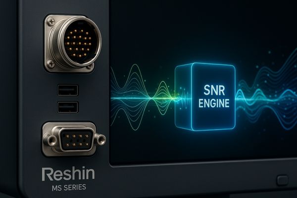
At Reshin, we integrate a suite of specific technologies into our medical displays, such as models in our MS series (surgical displays) and MD series (diagnostic displays), to ensure optimal signal integrity and image quality. Our proprietary Signal-to-Noise Ratio (SNR) enhancement engines13 are a core component. These systems, often a sophisticated blend of hardware and intelligent software algorithms, work to maximize the strength of the desired video signal relative to the underlying noise floor. This can involve input signal conditioning to amplify and clean the signal as it enters the display, along with advanced, adaptive filtering that targets noise without degrading critical image information. We also implement digital cross-talk reduction algorithms14. Cross-talk occurs when signals from adjacent wires or traces on a circuit board interfere with each other. This can cause ghosting or faint superimposed images. Our digital algorithms actively analyze and compensate for such interference, leading to a sharper and cleaner final image. Furthermore, the physical interfaces are critical. We utilize industrial-grade, robustly shielded connectors15. These connectors provide secure mechanical connections and feature comprehensive metal shielding that ensures excellent electrical continuity with the cable shield. This minimizes the risk of EMI entering or exiting the display at these vulnerable points. This holistic approach, combining advanced internal processing with robust physical design, allows our displays, including the MD120C diagnostic monitor, to deliver stable, clear, and reliable imaging performance, even when surrounded by the multitude of electronic devices found in modern operating rooms and radiology suites.
Conclusion
Medical displays employ robust hardware, intelligent algorithms, and effective shielding. These methods work synergistically to deliver the clear, noise-free images essential for accurate medical diagnosis and effective treatment. To learn more about Reshin’s high-performance, noise-free medical displays, contact us at martin@reshinmonitors.com.
-
Learn about the techniques hospitals use to maintain a clean signal, vital for high-quality medical imaging. ↩
-
Explore advancements in medical display technologies that enhance image clarity, crucial for accurate diagnoses. ↩
-
Discover the methods that lead to pristine images in medical imaging, essential for effective patient care. ↩
-
Understanding EMI is crucial for improving medical display quality and ensuring accurate diagnostics. Explore this link for in-depth insights. ↩
-
RFI can significantly affect medical imaging quality. Learn more about its effects and solutions to mitigate it. ↩
-
Understanding low-noise power supplies can enhance your knowledge of how medical displays maintain image quality and reliability. ↩
-
Exploring differential signal transmission will reveal how it helps in reducing noise and improving signal integrity in medical displays. ↩
-
Learning about internal signal equalizers will provide insights into how they enhance signal quality in medical displays, crucial for accurate imaging. ↩
-
Understanding how to effectively reduce image noise can significantly enhance the clarity of medical displays, improving diagnostic accuracy. ↩
-
Understanding shielding can enhance your knowledge of protecting electronic signals from interference, crucial for reliable performance. ↩
-
Learning about Faraday cages can provide insights into effective methods for shielding against electromagnetic interference. ↩
-
Exploring grounding will help you grasp its role in ensuring safety and signal integrity in electronic devices. ↩
-
Understanding SNR enhancement engines can help you appreciate their role in improving image quality in medical displays. ↩
-
Explore how these algorithms enhance image clarity by reducing interference, crucial for medical imaging applications. ↩
-
Learn about the importance of these connectors in ensuring reliable connections and minimizing EMI in medical displays. ↩


