Radiologists often ask me if the leap from a 2MP to a 5MP monitor truly impacts their diagnostic capabilities in mammography. When early cancer detection hangs in the balance, this question is critical. So, does the resolution of a mammography monitor genuinely make a difference?
Yes, the difference between 2MP and 5MP in mammography monitors significantly matters. A 5MP resolution is crucial for detecting subtle microcalcifications and lesions that might be missed on 2MP displays, directly impacting diagnostic accuracy in breast cancer screening.
The choice of display resolution for mammography is more than a technical detail. It is a fundamental factor that can influence patient outcomes. In this article, I will explore why higher resolution is so important in breast imaging. We will look at how resolution affects the ability to diagnose with accuracy and confidence.
What’s the difference between 2MP and 5MP resolution in display terms?
The terms "2MP" and "5MP" can seem like just numbers. Technical specifications often create confusion. What do these megapixel ratings actually mean for the clarity of images in a mammography reading room?
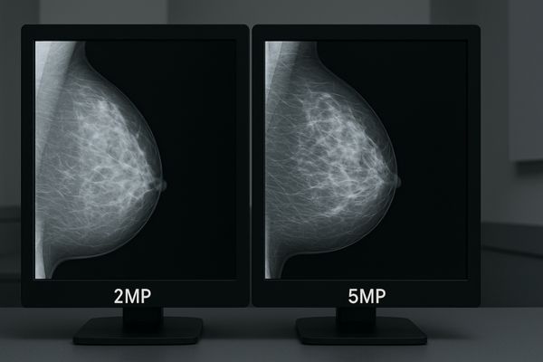
In display terms, 5MP resolution provides approximately 5 million pixels, while 2MP offers around 2 million pixels. This superior pixel density in 5MP monitors allows for the display of much finer details and sharper images, which is essential for medical diagnostics.
![]()
When we talk about "MP" or megapixels in the context of a display, we are referring to the total number of individual dots, or pixels, that make up the image on the screen. A 2-megapixel monitor1, commonly with a resolution like 1920×1080 (Full HD) or 1600×1200, has roughly two million pixels. In contrast, a 5-megapixel monitor2, typically featuring a resolution around 2560×2048 (often portrait for mammography), boasts over five million pixels. This substantial increase in pixel count, especially when applied to a similar physical screen size, results in a much higher pixel density. Higher pixel density means that each pixel is smaller and more closely packed, allowing the display to render finer details with greater sharpness and clarity. While 2MP monitors are adequate for many general radiology tasks where findings might be larger or more distinct, mammography demands the visualization of extremely subtle structures. The intricate patterns of breast tissue, the minute signs of early disease, such as tiny microcalcifications or faint lesion borders, require the enhanced detail that only a higher pixel density3 can provide. This difference is fundamental to ensuring that radiologists have the best possible visual information for their critical interpretations.
2MP vs. 5MP: A Quick Comparison
To better understand the practical differences, let us consider a direct comparison. The following table outlines key distinctions:
| Feature | 2MP Monitor | 5MP Monitor |
|---|---|---|
| Total Pixels | Approx. 1.9 to 2.1 million (e.g., 1600×1200 or 1920×1080) | Approx. 5.2 million (e.g., 2048×2560) |
| Typical Use | General radiology, clinical review, non-diagnostic | Primary diagnostic mammography |
| Detail Level | Standard, suitable for larger structures | Excellent, crucial for minute, subtle details |
| Pixel Density | Lower on a comparable screen size | Higher on a comparable screen size |
| Image Fidelity | May require scaling for full mammography dataset | Often allows 1:1 pixel mapping of image data |
This table highlights why 5MP resolution is specifically recommended for tasks where the smallest details are of utmost importance.
Why does mammography require higher resolution than general radiology?
Radiologists examine a wide variety of medical images. Some abnormalities are easier to spot than others. Why does mammography specifically necessitate a higher display resolution compared to other radiological studies?
Mammography requires higher resolution because critical diagnostic signs, like microcalcifications or the subtle edges of spiculated masses indicating early cancers, are extremely small. Insufficient resolution risks these vital details being overlooked, especially in dense breast tissue.
The unique challenge of mammography lies in the subtlety and minuteness of the earliest signs of breast cancer. Microcalcifications4, which can be an early indicator of ductal carcinoma in situ (DCIS) or invasive cancer, are tiny deposits of calcium. These can be smaller than 0.5 millimeters. To reliably detect, count, and characterize the morphology (shape, distribution) of these microcalcifications, a monitor must be able to resolve these very fine details. Similarly, early-stage tumors, particularly spiculated masses5, may present with very subtle architectural distortions or faint tendrils extending into the surrounding tissue. These subtle spiculations are crucial for diagnosis but can be easily obscured or averaged out by displays with lower pixel density. The challenge is further amplified in women with dense breast tissue6. Dense tissue can mask underlying lesions, making it even more critical that the display technology provides the maximum possible detail and contrast. General radiology often deals with larger anatomical structures or pathologies with higher intrinsic contrast. While clarity is always important, the sheer tininess of key findings in mammography places a unique and non-negotiable demand for superior resolution. Missing these early signs due to inadequate display capabilities can lead to delayed diagnosis, potentially impacting treatment options and patient prognosis.
How does resolution affect microcalcification and lesion detection?
Those tiny specks or faint patterns on a mammogram can be the earliest signs of cancer. Their visibility is paramount for a radiologist. How precisely does a monitor’s resolution influence the ability to detect these critical findings?
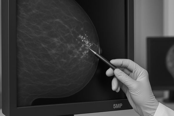
Higher resolution, such as that found in 5MP monitors, allows for 1:1 pixel mapping of digital mammography images. This avoids scaling or interpolation, preserving the integrity of fine details, thus making microcalcifications and subtle lesion margins clearer and easier to detect accurately.
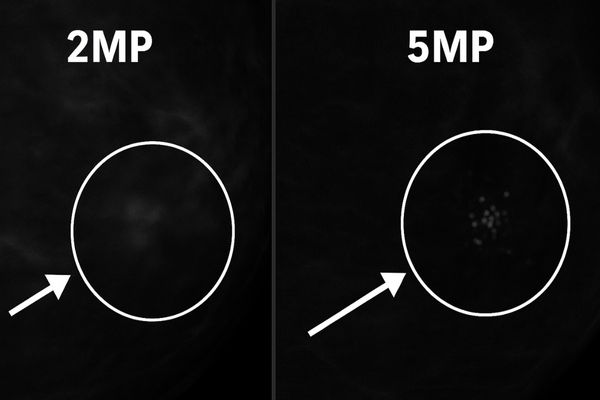
The impact of resolution on detecting microcalcifications7 and subtle lesions is direct and significant. Digital mammography images are captured at a certain intrinsic resolution. Ideally, the display monitor should have enough pixels to show the image data on a one-to-one basis, meaning each pixel in the acquired image corresponds to at least one pixel on the display. This is known as 1:1 pixel mapping8. When a lower-resolution monitor, such as a 2MP display, is used to view a high-resolution mammography image, the monitor does not have enough pixels to display all the image data natively. Consequently, the image must be downscaled or an interpolated (averaged) version must be shown, especially when viewing the image at its full size or "true size." This process of scaling or interpolation can blur or even eliminate the finest details, such as individual microcalcifications or the delicate edges of a lesion. With a 5MP monitor, such as our MD52G 5MP Grayscale Mammography Monitor, there are enough pixels to display the mammographic image much closer to, or at, a 1:1 pixel mapping. This preserves the sharpness and integrity of the original image data. Radiologists can then more clearly discern the presence, morphology, and distribution of microcalcifications, which are critical factors in differentiating benign from malignant clusters. The American College of Radiology (ACR) and DICOM (Digital Imaging and Communications in Medicine) standards recognize this, recommending 5MP monitors9 for primary diagnostic interpretation in mammography to ensure diagnostic integrity.
Are there trade-offs in brightness, speed, or cost between 2MP and 5MP monitors?
Adopting advanced technology often involves evaluating certain compromises. Higher performance can sometimes mean higher costs or other considerations. What are the practical trade-offs when a facility is choosing between 2MP and 5MP mammography displays?
Yes, 5MP mammography monitors are generally more expensive than 2MP displays. They also require more sophisticated electronics for precise brightness control and consistent image processing. However, these are considered necessary investments for the diagnostic accuracy required in breast screening.

There are indeed trade-offs to consider when comparing 2MP and 5MP mammography monitors10, primarily concerning cost and the technology required to drive them. Manufacturing high-resolution 5MP panels with the exceptional brightness and uniformity demanded by mammography is more complex and, therefore, more expensive than producing 2MP panels. The typical luminance required for mammography reading is significantly higher than for general radiology, often exceeding 500 cd/m² and ideally reaching towards 1000 cd/m² to perceive subtle contrast differences in dense tissue. Achieving and maintaining such high brightness levels11 consistently across a large 5MP screen, while also ensuring longevity, demands advanced backlight technology and meticulous quality control. Furthermore, the electronics needed to process and render over five million pixels with precise grayscale accuracy, adhering to the DICOM GSDF standard12, are more sophisticated. This contributes to the higher overall cost of 5MP systems. While refresh rates and response times (speed) are generally sufficient for static mammography images on both types, the sheer amount of data being handled by a 5MP display means that the paired graphics card also needs to be capable. Despite the higher initial investment, the clinical benefits of improved diagnostic accuracy and the potential for earlier cancer detection with 5MP monitors typically outweigh the cost considerations for dedicated breast imaging centers. It is an investment in patient care and diagnostic confidence.
How does Reshin address diagnostic performance in mammography displays?
Choosing the right medical display is a cornerstone of excellent patient care. Radiologists must have complete confidence in their diagnostic tools. How do our mammography displays at Reshin ensure they deliver top-tier performance for this critical task?
Reshin addresses diagnostic performance with specialized mammography monitors, like our MD50C 5MP Color Mammography Monitor. These feature high-luminance 5MP panels, precise DICOM Part 14 calibration, integrated brightness stabilization, and exceptionally accurate grayscale rendering, all tailored for demanding breast imaging environments.
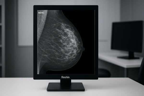
At Reshin, we understand the critical demands of mammography. We design our mammography-specific displays, such as the MD50C 5MP Color Mammography Monitor13 and the MD52G 5MP Grayscale Mammography Monitor, with features directly aimed at maximizing diagnostic performance. These monitors utilize high-quality 5MP resolution panels capable of achieving the high luminance levels (often exceeding 1000 cd/m²) essential for perceiving subtle density variations in breast tissue. Crucially, each monitor undergoes meticulous factory calibration to the DICOM Part 14 Grayscale Standard Display Function14 (GSDF). This ensures that images are displayed with perceptually linearized grayscale, meaning that differences in pixel values translate into discernible differences in brightness consistently. To maintain this performance over time, our monitors incorporate brightness stabilization systems15. These often use built-in front sensors that continuously measure luminance and automatically adjust the backlight to compensate for warm-up drift and long-term aging, ensuring consistent image quality day after day. Precise grayscale rendering, with the ability to display a multitude of distinct shades of gray, is fundamental for differentiating subtle tissue characteristics. We also incorporate features like ambient light sensors and user-friendly quality assurance software to help maintain optimal viewing conditions and compliance with medical imaging standards. These combined technologies ensure that our mammography displays provide the clarity, consistency, and reliability that radiologists need for confident diagnoses.
Conclusion
In the specialized field of mammography, selecting a 5MP resolution monitor is not a luxury; it is a clinical necessity. This higher resolution directly enhances the detection of subtle, early signs of cancer, significantly improving diagnostic accuracy and, ultimately, patient outcomes. To ensure your mammography practice is equipped with high-resolution diagnostic displays, contact Reshin at martin@reshinmonitors.com.
-
Exploring the benefits of a 2-megapixel monitor can help you understand its applications in general radiology tasks. ↩
-
Discover how a 5-megapixel monitor enhances mammography, providing vital detail for early disease detection. ↩
-
Understanding pixel density is crucial for evaluating display quality, especially in fields like radiology where detail is paramount. ↩
-
Understanding microcalcifications is crucial for early breast cancer detection. Explore this link to learn more about their role in mammography. ↩
-
Spiculated masses can indicate serious conditions. Discover their importance in breast imaging and diagnosis by exploring this resource. ↩
-
Dense breast tissue can obscure lesions. Learn how it impacts mammography and what to do about it by checking this informative link. ↩
-
Microcalcifications can indicate early signs of breast cancer; learning more can improve understanding of their significance in diagnostics. ↩
-
Understanding 1:1 pixel mapping is crucial for ensuring accurate image interpretation in mammography, enhancing diagnostic accuracy. ↩
-
5MP monitors are essential for clear image display, allowing radiologists to detect critical details in mammograms effectively. ↩
-
Explore the benefits of 5MP mammography monitors, including improved diagnostic accuracy and earlier cancer detection, crucial for patient care. ↩
-
Learn why high brightness levels are critical for perceiving subtle contrast differences in dense tissue, vital for accurate diagnoses. ↩
-
Understanding the DICOM GSDF standard is essential for ensuring accurate grayscale rendering in medical imaging, enhancing diagnostic quality. ↩
-
Explore the advantages of 5MP Color Mammography Monitors for enhanced diagnostic accuracy and image quality in mammography. ↩
-
Understanding DICOM Part 14 is crucial for ensuring accurate grayscale rendering in medical imaging, enhancing diagnostic confidence. ↩
-
Learn how brightness stabilization systems maintain consistent image quality, crucial for reliable diagnostics in medical imaging. ↩