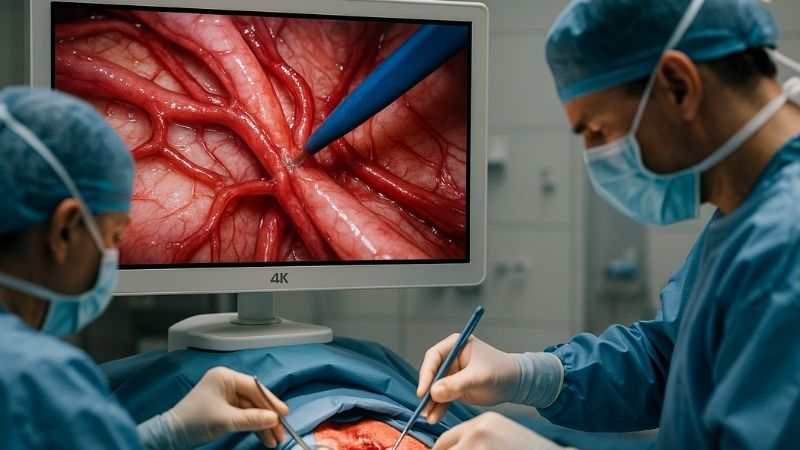
Vascular surgeons often grapple with visualizing minute blood vessels and intricate anatomical structures during delicate procedures. Inadequate image resolution can obscure crucial details, potentially leading to complications or suboptimal outcomes. Imagine the stress of trying to identify a tiny vessel or place a precise suture when your monitor doesn’t offer the clarity needed. This lack of detail can increase procedural time and risk. High-resolution monitors, particularly 4K, offer the solution by providing unparalleled image clarity, enabling surgeons to confidently navigate complex vascular networks.
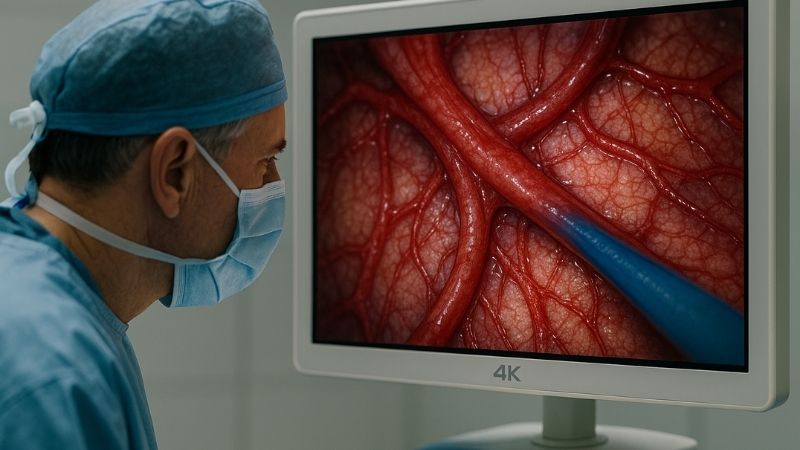
For vascular surgery, 4K Ultra HD (3840×2160 pixels) resolution is generally considered optimal. This high pixel density provides exceptional detail, allowing surgeons to clearly visualize fine vessels, subtle tissue changes, and the precise placement of sutures or stents. This clarity is paramount for enhancing surgical accuracy and patient safety in complex vascular interventions.
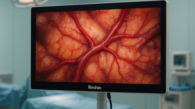
As someone who has been deeply involved in developing surgical display technology at Reshin, I understand the critical role resolution plays. Let’s explore why 4K stands out for vascular applications.
Why is image resolution so critical in vascular surgery?
Vascular procedures often involve tiny, delicate structures where misinterpretation can have serious consequences. Poor resolution can hide these critical details, potentially leading to errors. High resolution, however, reveals these nuances, significantly enhancing safety and precision.
Image resolution is paramount in vascular surgery because it directly impacts the surgeon’s ability to discern fine anatomical details, crucial for navigating complex vessel networks and minimizing iatrogenic injury.
"Vascular surgery requires identifying extremely small vessels, particularly in endoscopic or microsurgical procedures. High-resolution monitors significantly enhance anatomical detail perception1, reducing procedural risk." My experience at Reshin, working alongside surgeons and understanding their needs, has consistently shown that those performing these intricate tasks rely heavily on the visual information presented. Think about dissecting a small arteriole or placing a micro-suture; the difference between seeing a clear, sharp edge versus a slightly blurred one can be the difference between a successful anastomosis2 and a potential leak. High resolution translates to more visual data points per inch, allowing for a more accurate and faithful representation of the surgical field. This isn’t just about seeing better; it’s about understanding the tissue interactions, identifying subtle pathological changes, and executing maneuvers with greater confidence and precision. The clarity afforded by higher resolutions minimizes ambiguity, which is absolutely critical when dealing with structures where millimeters, or even micrometers, matter immensely.
What clinical benefits does 4K resolution offer in vascular procedures?
Surgeons need to discern subtle changes in vessel walls, plaque characteristics, or the presence of minute thrombi. FHD might not consistently reveal these early or subtle signs, potentially impacting critical intraoperative decisions. 4K resolution, with its superior detail, provides the clarity needed to detect these important nuances.
4K resolution offers significant clinical benefits, including enhanced visualization of subtle tissue differentiation, improved depth perception due to clearer textures, and more precise instrument guidance, leading to potentially better patient outcomes.
"Compared to 1080p (FHD), 4K resolution3 (3840×2160) delivers four times the pixel density, allowing for clearer detail—critical for assessing vessel paths and suture points." This four-fold increase in pixels isn’t just a technical specification; it translates directly to a more detailed, richer, and more lifelike image on the screen. For vascular surgeons4, this means they can more accurately assess the morphology of a vessel, identify the extent and characteristics of atherosclerotic plaque, or distinguish between healthy and diseased tissue with greater certainty. When performing an endarterectomy, for example, the ability to clearly see the cleavage plane for plaque removal is vital. 4K resolution allows for this level of detail, making it easier to remove the plaque thoroughly without damaging the delicate vessel wall. Similarly, when placing stents or grafts, the precise visualization of the vessel lumen and the device itself, as offered by monitors like the Reshin MS321PB, ensures accurate deployment and optimal apposition against the vessel wall, which are key factors for long-term patency and procedural success.
How do FHD and UHD monitors differ in minimally invasive vascular operations?
Minimally invasive vascular surgery, like endovascular procedures, relies entirely on mediated vision through screens. FHD might not provide sufficient detail for these intricate, remotely performed tasks, potentially limiting precision. UHD, however, offers superior clarity essential for navigating complex anatomies.
In minimally invasive vascular operations, UHD (4K) monitors provide vastly superior image clarity and detail compared to FHD, crucial for navigating intricate anatomies and manipulating fine instruments with high accuracy.
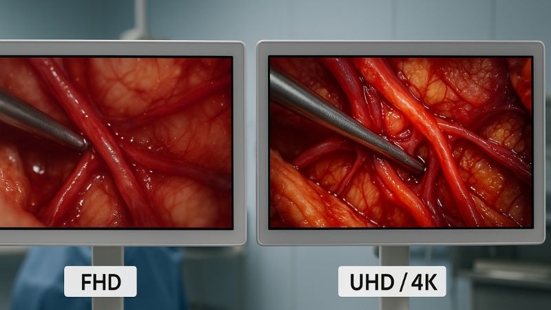
Minimally invasive vascular operations, such as endovascular aneurysm repair (EVAR)5, peripheral angioplasty, or thrombectomy, are performed using catheters and guidewires navigated under real-time imaging guidance, typically fluoroscopy or intravascular ultrasound (IVUS). The difference between Full HD (FHD) with 1920×1080 pixels and Ultra HD (UHD/4K) with 3840×2160 pixels in these scenarios is stark and impactful. While FHD offers a decent image, UHD provides four times the number of pixels. This massive increase in pixel density means that UHD monitors can display significantly more intricate detail from the imaging source. "UHD monitors6 paired with uncompressed digital video transmission reduce latency and image blur, which is essential for time-sensitive surgeries like aneurysm clipping or bypass grafting," and indeed for precise catheter manipulation7. For a surgeon guiding a catheter through a tortuous iliac artery or deploying a stent in a coronary vessel, this translates to a clearer depiction of the vessel’s course, easier identification of crucial side branches, and a more accurate assessment of lesion characteristics like stenosis severity or plaque morphology. The sharper, more detailed image reduces eye strain and allows for more confident and precise manipulation of instruments, which is absolutely critical when working in delicate and often diseased vascular territories.
How do Reshin surgical monitor resolutions meet the needs of vascular surgery?
Vascular surgeons require displays that are not only high-resolution but also consistently reliable under demanding OR conditions. Using subpar monitors can directly compromise surgical precision and, ultimately, patient outcomes. Reshin addresses this by offering specialized monitors engineered for these exacting demands.
Reshin surgical monitors, particularly our 4K models, are engineered with high pixel density, exceptional brightness, robust color fidelity, and low latency to meet vascular surgery’s stringent visual demands effectively.
At Reshin, we’ve poured years of R&D into understanding the specific visual needs of different surgical specialties, and vascular surgery has always been a key focus. The feedback from leading vascular surgeons has been unequivocal: resolution, color accuracy8, and reliability are paramount. "Models like the Reshin MS321PB and MS430PC offer native 4K resolution with high brightness, anti-glare glass, and precise color calibration—perfectly suited for vascular imaging demands." These features are not just specifications on a datasheet; they translate into tangible benefits in the operating room. The native 4K resolution9 ensures that the image captured by high-definition endoscopes, surgical microscopes, or angiography systems is displayed with maximum fidelity, without downscaling or loss of detail. High brightness and effective anti-glare properties10 ensure clear visibility even under the bright, often variable, OR lighting conditions. Precise color calibration helps in differentiating subtle tissue variations, crucial for identifying perfusion, ischemia, or the health of vessel walls. Our monitors are designed to deliver this, empowering vascular surgeons with the visual confidence they need.
Does resolution affect the precision of vascular anatomical visualization?
Precise anatomical visualization forms the bedrock of successful vascular surgery. If critical details are obscured by lower resolutions, the surgeon’s ability to operate with pinpoint accuracy is compromised. Higher resolution directly and significantly enhances this visualization precision.
Yes, resolution profoundly affects the precision of vascular anatomical visualization. Higher resolutions enable surgeons to discern finer structures and subtle textural differences, improving accuracy in diagnosis and intervention.
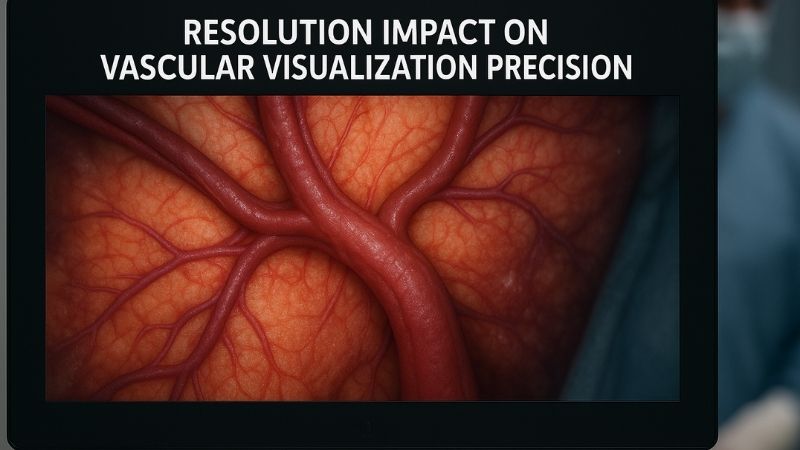
Absolutely, resolution has a direct and significant impact on the precision of vascular anatomical visualization11. Consider the task of identifying and preserving tiny perforator vessels during a complex bypass procedure, accurately placing a fenestration in a custom-made stent graft to align perfectly with a visceral artery, or discerning the subtle web-like structures of a vascular malformation. These tasks demand an exceptional level of detail that lower resolutions simply cannot provide. Higher resolution means more pixels per inch, which translates to a sharper, more detailed, and more true-to-life image. This allows surgeons to see finer structures12, subtle variations in tissue texture, and the exact spatial relationship between anatomical landmarks and their instruments. "While high resolution is an advantage, it must be paired with proper screen size, viewing distance, and signal quality to fully realize its clinical value." This is a crucial point we always emphasize. A 4K monitor, such as our Reshin MS430PC, when integrated into a complete 4K imaging chain13 (camera, processor, cabling) and viewed at an appropriate distance, allows surgeons to appreciate the depth, detail, and subtle visual cues that guide precise surgical actions. This enhanced precision can lead to reduced operative time, fewer iatrogenic injuries, and ultimately, better, more durable patient outcomes.
Conclusion
Optimal resolution, ideally 4K UHD, is crucial in vascular surgery. It enhances anatomical detail, improves procedural precision, and supports superior patient outcomes through consistently clearer visualization. To discover high-resolution displays designed for vascular procedures, contact Reshin at martin@reshinmonitors.com.
-
Understanding anatomical detail perception can lead to improved surgical techniques and better patient outcomes. ↩
-
Learn about the critical factors that ensure successful anastomosis, which is vital for patient recovery and surgical success. ↩
-
Explore how 4K resolution enhances medical imaging, providing clearer details crucial for surgical precision and patient outcomes. ↩
-
Learn about the advanced techniques vascular surgeons employ, including the importance of imaging in successful procedures. ↩
-
Explore this resource to understand the intricacies of EVAR, a key procedure in minimally invasive vascular surgery. ↩
-
Discover how UHD monitors enhance surgical precision and reduce latency, crucial for successful outcomes in vascular procedures. ↩
-
Learn about effective catheter manipulation techniques that can significantly improve surgical results and patient safety. ↩
-
Understanding the significance of color accuracy can improve surgical outcomes by aiding in the identification of tissue variations. ↩
-
Explore how native 4K resolution enhances surgical imaging, providing clarity and detail crucial for successful procedures. ↩
-
Learn how anti-glare technology enhances visibility in bright OR conditions, ensuring surgeons can perform with precision. ↩
-
Understanding vascular anatomical visualization is crucial for improving surgical precision and patient outcomes. Explore this resource to learn more. ↩
-
Explore techniques and technologies that enable surgeons to visualize finer structures, enhancing surgical accuracy and safety. ↩
-
Discover how a 4K imaging chain can significantly improve surgical detail and precision, leading to better patient care. ↩