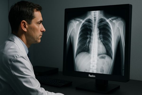
Difficulty in spotting faint details on medical scans can lead to diagnostic uncertainty and added stress for clinicians. Missing subtle lesions due to poor display contrast risks patient safety and undermines diagnostic confidence. High contrast ratio displays address this challenge by making the invisible visible.
The top five benefits of high contrast ratios in medical imaging are:
1.enhanced visibility of subtle anatomical structures.
2.improved diagnostic accuracy in low-contrast areas.
3.reduced eye strain for radiologists.
4.better differentiation between healthy and abnormal tissue.
5.and critical support for demanding modalities like mammography and MRI.
These advantages collectively lead to higher diagnostic confidence, workflow efficiency, and better patient outcomes.
Let’s delve deeper into how these benefits translate into tangible improvements in the daily practice of diagnostic and surgical imaging.
Enhanced Visibility of Subtle Anatomical Structures
When viewing medical images, faint anatomical structures can easily blend into the background on a standard display. This lack of clear demarcation creates ambiguity and can impede an accurate assessment.
A high contrast ratio is crucial because it amplifies the distinction between different shades of gray, effectively pulling fine details like tiny blood vessels, subtle fractures, or faint tissue planes into focus.
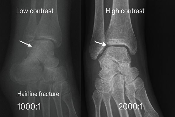
In my years leading Reshin, I’ve learned that a high contrast ratio1 can mean the difference between detecting a faint lesion and missing it entirely. Contrast ratio is the fundamental measure of a display’s ability to distinguish between its brightest white and its darkest black. A monitor with a high static contrast ratio—the inherent contrast of the panel itself—can render a wider gamut of distinct shades between these two extremes. For example, a consumer-grade monitor might have a 1000:1 ratio, but a specialized medical display like our Reshin MD33G 3MP diagnostic monitor features a contrast ratio of 2000:1. This isn’t just a number; it means the display can resolve more Just Noticeable Differences (JNDs)2, allowing a radiologist to perceive subtle changes in tissue density that would be invisible on a lesser screen. I remember a case shared by a partner hospital where a hairline fracture in a trauma patient’s X-ray was initially missed on a standard review station but was immediately obvious on one of our high-contrast displays3. That experience solidified my belief that we are not just selling monitors; we are providing certainty.
| Display Type | Typical Static Contrast Ratio | Visibility of Subtle Structures | Clinical Implication |
|---|---|---|---|
| Standard Consumer Monitor | 1000:1 | Low; faint details merge with background | Potential for missed findings |
| Professional Graphics Monitor | 1200:1 | Moderate; improved detail but not DICOM-calibrated | Risk of inaccurate grayscale representation |
| Reshin Medical Display | 1500:1 – 2000:1+ | High; excellent separation of gray shades | Enhanced detection of subtle pathologies |
Improved Diagnostic Accuracy in Low-Contrast Areas
Many diagnostic challenges lie in low-contrast regions, where different soft tissues have very similar radiographic densities. On a subpar display, these areas can appear as a uniform, undifferentiated mass, hiding critical pathology.
In areas with inherently low contrast, such as the brain’s gray and white matter or soft tissue in the abdomen, a high contrast ratio allows for superior differentiation, directly improving diagnostic accuracy.
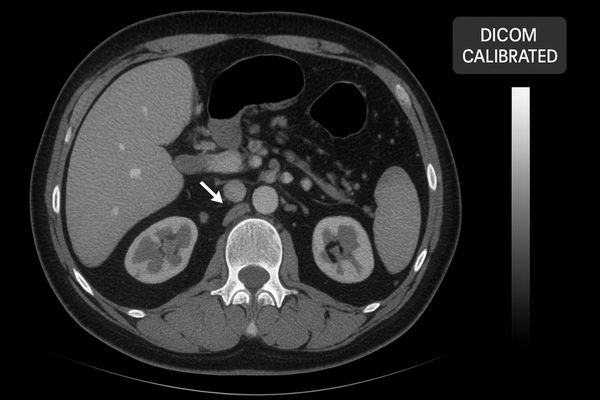
As I’ve often said, in radiology, clarity isn’t just about resolution—it’s about contrast. You can have a monitor with millions of pixels, but if it can’t distinguish between subtle differences in gray, those pixels aren’t conveying the necessary information. This is particularly true for low-contrast studies, like CT scans of the abdomen or non-contrast brain MRIs. In these images, the difference in attenuation between healthy tissue and an early-stage infiltration can be incredibly slight. A high-contrast monitor4, factory-calibrated to the DICOM Part 14 standard5, ensures a perceptually linear grayscale response6. This means that every single increase in the image data’s grayscale value corresponds to a visually distinct increase in brightness on the screen. This capability is engineered into every Reshin display, with stabilization sensors ensuring that a radiologist can confidently detect subtle edema or a nascent tumor within what would otherwise look like homogenous tissue. This technology directly empowers clinicians to make more definitive diagnoses earlier in the disease process.
Reduced Eye Strain for Radiologists During Long Sessions
Radiologists spend hours reading hundreds of complex images daily, a task that places immense strain on their eyes. This visual fatigue is a hidden threat that can degrade focus and increase the potential for perceptual errors.
A display with a high, stable contrast ratio presents a clearer, more defined image that is easier for the brain to process, thereby reducing the cognitive load and physical eye strain on the reader.
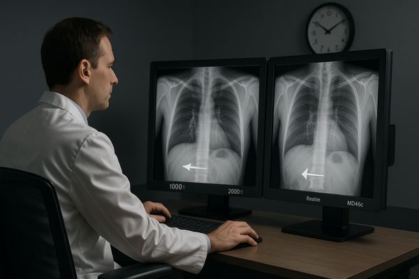
Eye fatigue is a critical factor in diagnostic imaging, and it’s an area where display technology can make a significant impact. When a radiologist has to consciously or subconsciously struggle to differentiate shades of gray, their visual system works overtime. This leads to tired eyes, headaches, and a decline in cognitive performance over a long reading session, which can increase the risk of errors. A high-contrast display mitigates this by making the information on the screen unambiguous. The separation between different tissues is crisp and clear, allowing for quicker and more effortless recognition. This means less time is spent simply trying to see the image and more time is available for analyzing the pathology. At Reshin, our commitment extends beyond image fidelity to the well-being of the clinician. Our diagnostic displays, like the dual-screen MD46C, are engineered with high contrast and integrated ambient light sensors. These features work together to maintain a consistent, comfortable viewing experience, supporting peak performance from the first read of the day to the last.
Better Differentiation Between Healthy and Abnormal Tissue
The core of many diagnoses, particularly in oncology, is distinguishing abnormal tissue from the healthy tissue surrounding it. If a display blurs the lines between them, it can lead to diagnostic uncertainty or misjudgment.
High contrast is essential for clearly delineating the margins of tumors, lesions, or other pathologies, providing the sharp visual boundaries needed for confident diagnosis, staging, and treatment planning.
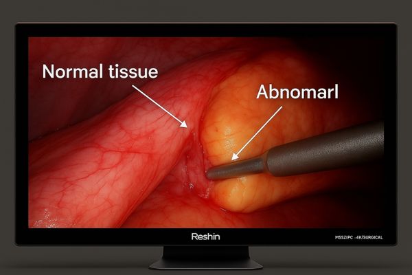
In oncology imaging, contrast can influence life-or-death decisions. This is not an exaggeration. The ability to precisely visualize the edges of a mass is critical for determining whether it is benign or malignant, assessing its size, and seeing if it has invaded surrounding structures. This clarity is crucial not only for radiologists but also for surgeons. During a minimally invasive procedure7, a surgeon relies entirely on the monitor to guide their instruments. I’ve spoken with partners like Alex Müller from Mediview Technologies in Germany, who develop endoscopic systems. His clients, the surgeons, demand displays with zero lag and exceptional contrast because they need to differentiate between delicate tissue planes in real-time. A display like our Reshin MS321PC 4K surgical monitor8 provides the high contrast9 necessary to see these subtle differences, ensuring safer and more precise surgery. Without it, determining the exact extent of a tumor becomes a matter of approximation, which is unacceptable in modern medicine.
| Contrast Level | Impact on Tissue Differentiation | Surgical/Treatment Planning Implication |
|---|---|---|
| Low Contrast | Blurry or indistinct margins between tissues | High uncertainty; risk of incomplete tumor resection |
| Medium Contrast | Margins are visible but may lack fine detail | Better, but may still miss subtle satellite lesions or invasion |
| High Contrast | Crisp, well-defined boundaries and internal texture | High confidence for accurate measurements and clean surgical margins |
Critical Support for Modalities Like Mammography and MRI
Modern imaging modalities like digital mammography and MRI produce incredibly rich and complex datasets. However, this data often contains crucial information in the form of very subtle, low-contrast variations that standard displays cannot render.
For modalities that live and die by subtle detail—like detecting microcalcifications in mammograms or soft tissue lesions in MRI—a high-contrast display is not just beneficial, it’s a fundamental requirement.
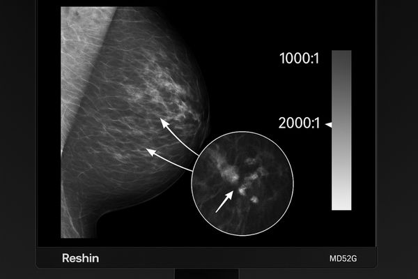
Modern imaging techniques demand displays that can match their output fidelity. The imaging chain is only as strong as its weakest link, and very often, that link is the display. Take digital breast tomosynthesis (DBT)10. The primary goal is often to detect tiny, bright specks known as microcalcifications against a background of dense breast tissue. This requires an exceptionally high contrast ratio11 and luminance to make these specks "pop" visually. This is why specialized displays like our Reshin MD52G 5MP Grayscale Mammography Monitor are engineered with contrast ratios exceeding 1500:1 and high brightness levels. Similarly, an MRI’s wide dynamic range requires a capable display to show both the bright fat signals and dark fluid signals in the same image without losing detail. Multi-modality monitors like the Reshin MD120C 12MP display provide the flexibility to read images from various scanners on one workstation, ensuring that no detail is lost between the scanner and the radiologist’s eyes.
| Imaging Modality | Key Diagnostic Feature | Required Contrast Characteristic | Recommended Reshin Model |
|---|---|---|---|
| Mammography | Microcalcifications, subtle masses | Extremely high contrast and luminance | MD52G, MD50C |
| MRI (Soft Tissue) | Lesion characterization, edema | High contrast for subtle gray-level differences | 8MP Display, MD120C |
| CT (Lung) | Ground-glass opacities, nodules | High contrast to differentiate air and tissue | MD33G, MD26GA |
| Endoscopy | Tissue planes, vascularity | Real-time high contrast and color accuracy | MS275P, MS321PC |
Conclusion
Ultimately, a high contrast ratio is not a luxury feature but a core technological pillar of modern medical displays, directly enhancing diagnostic accuracy, improving clinician efficiency, and supporting better patient outcomes. To experience high-contrast medical displays engineered for clinical excellence, contact Reshin at martin@reshinmonitors.com.
-
Understanding contrast ratio is crucial for medical professionals to ensure accurate diagnosis and patient care. Explore this link to learn more. ↩
-
JNDs play a vital role in detecting subtle changes in medical images. Discover how they impact diagnostic accuracy. ↩
-
High-contrast displays enhance image clarity, crucial for accurate diagnoses. Learn more about their advantages in medical settings. ↩
-
Understanding high-contrast monitors can enhance your knowledge of their critical role in radiology, leading to better diagnostic outcomes. ↩
-
Exploring the DICOM Part 14 standard will provide insights into how it ensures image quality and consistency in medical imaging. ↩
-
Learning about perceptually linear grayscale response can help you understand how it affects image interpretation and diagnosis accuracy. ↩
-
Discover how minimally invasive techniques improve recovery times and reduce complications for patients. This resource is invaluable. ↩
-
Learn about the essential features of surgical monitors that can significantly impact surgical outcomes and patient safety. ↩
-
Understanding high contrast in imaging can enhance surgical precision and patient outcomes. Explore this link to learn more. ↩
-
Explore the advantages of DBT in detecting microcalcifications and improving breast cancer screening accuracy. ↩
-
Understanding contrast ratio is crucial for optimizing image quality in medical displays, enhancing diagnostic accuracy. ↩