Relying on displays that cannot render fine details means critical information is lost. This technical limitation directly compromises diagnostic accuracy and can negatively affect patient care decisions.
The evolution of detail reproduction in medical displays extends beyond mere pixel count. It now encompasses a sophisticated interplay of grayscale precision, luminance control, and intelligent image processing algorithms to achieve superior diagnostic clarity.
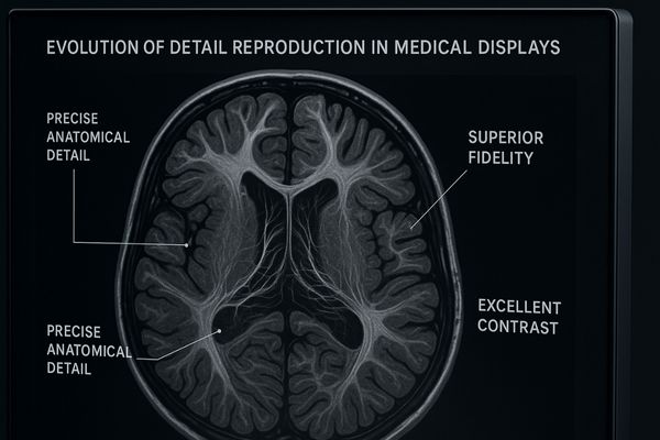
For decades, the quality of a medical display was judged primarily by its resolution. While the number of pixels is important, it is only one part of a much larger story. True detail reproduction1—the ability to show the most subtle variations in tissue density or the faintest outlines of a lesion—is a far more complex challenge. It requires a harmony between the physical screen, the electronics that drive it, and the software that processes the image data. As medical imaging modalities become more powerful and AI-driven tools2 enter clinical practice, the demands on display technology have intensified. This article explores the technological evolution that has enabled modern displays to render medical images with unprecedented fidelity.
Understanding Detail Reproduction in Medical Imaging
Thinking that more pixels automatically means more detail is a common oversimplification. This mindset leads to poor technology choices that fail to improve diagnostic capabilities.
Effective detail reproduction is not just about resolution. It is a function of the display’s ability to maintain grayscale linearity, luminance uniformity, and optimized image processing, all working together to present a clear and accurate image.
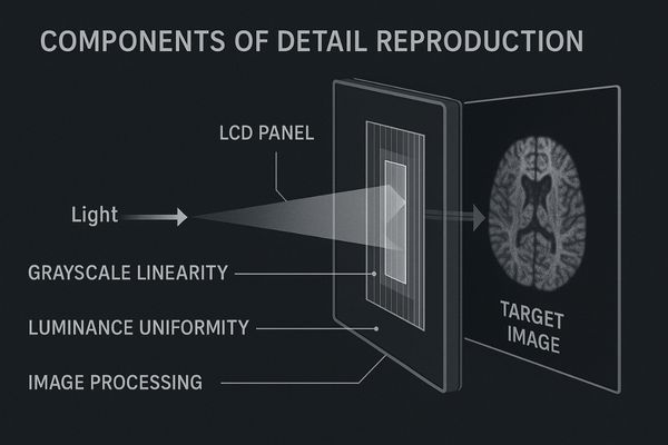
I have always emphasized that detail reproduction is a multi-faceted science. It is not something you can achieve by simply increasing the pixel count on a screen. The first critical element is grayscale linearity3, which is the display’s ability to show distinct, perceptible steps between different shades of gray. This is governed by the DICOM Part 14 standard and is essential for accurately representing different tissue densities. Another key factor is luminance uniformity4. The display must have consistent brightness across its entire surface, because any bright or dark patches can obscure or mimic pathology, leading to misinterpretation. Finally, intelligent algorithms play a crucial role. These internal processors optimize the incoming signal, reducing noise and sharpening details without introducing artificial artifacts. A display that excels in all these areas, such as the MD32C – 3MP Diagnostic Monitor, provides clinicians with a reliable and precise view, ensuring that the details captured by the scanner are faithfully delivered to the human eye.
| Component of Detail Reproduction | Why It Matters | Clinical Impact |
|---|---|---|
| Pixel Resolution | Defines the maximum level of spatial detail. | Ability to see small structures like microcalcifications. |
| Grayscale Linearity (DICOM) | Ensures accurate representation of tissue density. | Differentiating between subtle variations in soft tissue. |
| Luminance Uniformity | Prevents hot spots or dark areas from hiding information. | Consistent image quality across the entire screen. |
| Image Processing Algorithms | Reduces noise and enhances important features. | Improves clarity and reduces visual clutter in the image. |
Key Technologies Enhancing Image Precision
A standard display cannot render the subtle findings flagged by advanced AI software. This deficiency makes the sophisticated diagnostic tool less effective, wasting its full potential.
The precision of modern medical displays is enhanced by key technologies. These include high bit-depth processing for smoother grayscale transitions, integrated front sensors for continuous calibration, and advanced backlighting for superior contrast.
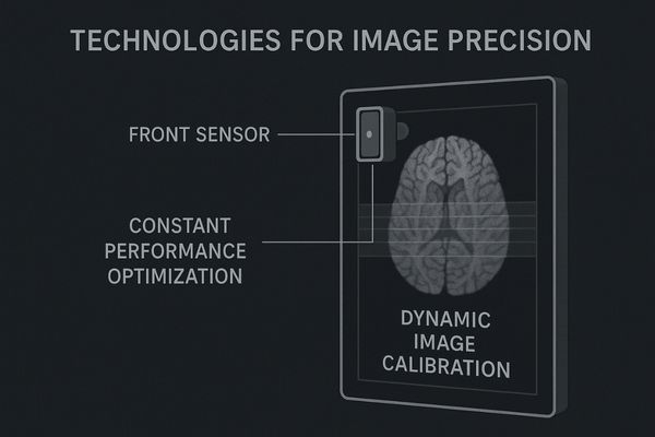
The rise of AI in diagnostics has created a new set of demands for medical displays. AI algorithms are incredibly skilled at detecting subtle patterns that a human might overlook, but this information is useless if the display cannot render it visibly. This has pushed manufacturers to develop technologies that enhance image precision at a granular level. One of the most important is higher bit-depth processing5. A standard display might use 8 bits to represent 256 shades of gray. A high-performance medical display uses 10-bit or even 12-bit processing, allowing for 1,024 or more shades. This creates a much smoother and more detailed grayscale, making low-contrast lesions more apparent. To maintain this precision over time, we integrate front sensors that constantly measure the screen’s output and automatically adjust it to stay compliant with the DICOM standard6. This ensures the display you use on day one is the same as the one you use years later. Our MD85CA is designed for these demanding applications, providing the bit depth and stability needed to serve as the perfect partner for AI-assisted diagnostics.
Milestones in the Evolution of Detail Rendering
Using older display technology is like trying to navigate with a faded, inaccurate map. Important clinical information appears washed out or is lost completely, leading to diagnostic uncertainty.
Over the last decade, the evolution of detail rendering has been marked by several milestones. The most significant include the adoption of High Dynamic Range (HDR), high bit-depth grayscale capabilities, and local dimming backlight technologies.
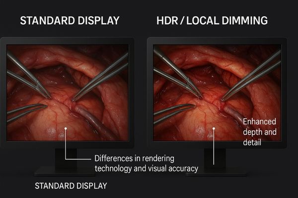
The journey to better detail reproduction has been marked by several key technological shifts. A decade ago, the focus was on improving the basic contrast ratio and brightness of LCD panels. The next major leap was the widespread adoption of high bit-depth processing, which dramatically increased the number of grayscale steps we could display. More recently, two technologies have had a profound impact. The first is High Dynamic Range (HDR)7. Borrowed from the consumer electronics world and adapted for medical use, HDR allows a display to show extremely bright and extremely dark areas in the same image without losing detail in either. This is critical in surgery, where a monitor must show deep, dark cavities alongside highly reflective surgical instruments. The second is local dimming8. This technology divides the LED backlight into multiple zones that can be independently brightened or dimmed. This significantly improves the contrast ratio, making blacks deeper and brights pop. The MS275P – 27" 4K Surgical Monitor incorporates these advancements, using HDR to give surgeons a clear, unambiguous view of the entire surgical field.
| Technology | Standard Dynamic Range (SDR) | High Dynamic Range (HDR) / Local Dimming |
|---|---|---|
| Contrast | Limited; blacks appear grayish. | High; deep blacks and bright highlights. |
| Detail in Shadows | Often crushed or lost. | Clearly visible and well-defined. |
| Detail in Highlights | Often clipped or washed out. | Retained without losing texture. |
| Overall Impression | Flat, less lifelike image. | Realistic, high-impact, and detailed image. |
Impact on Diagnostic Accuracy and Clinical Outcomes
When clinicians cannot trust what they see on their screen, they hesitate. This diagnostic uncertainty can lead to requests for additional scans, delayed treatment, and poorer patient outcomes.
The accurate reproduction of fine details has a direct and significant impact on diagnostic confidence. High-fidelity displays enable clinicians to detect and characterize subtle pathologies earlier and with greater certainty, which leads to improved clinical outcomes.
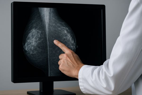
I have always believed that the ultimate measure of a medical display’s quality is its impact on a clinician’s confidence. All the technology we build into a monitor has one final purpose: to give a doctor the clearest possible view of the patient’s condition so they can make the best possible decision. When a display can accurately render the faint, spidery edges of an early-stage tumor or the subtle texture changes in soft tissue, it removes ambiguity. This has a powerful effect. It reduces diagnostic uncertainty9, which in turn can lead to fewer follow-up scans, faster treatment decisions, and ultimately, better patient outcomes. This is especially true in demanding fields like mammography10, where clinicians are searching for microcalcifications that can be as small as a single pixel. A display that fails to render these details faithfully fails the patient. That is why purpose-built monitors like the MD52G – 5MP Grayscale Mammography Monitor are so critical. They are engineered with the singular goal of maximizing detail reproduction to support the highest levels of diagnostic accuracy.
Future Innovations in Detail Reproduction
If we only focus on today’s display technology, we will be unprepared for the imaging tools of tomorrow. The rapid pace of innovation in medical imaging demands a constant, forward-looking approach to display design.
Future innovations in detail reproduction will be led by the broad adoption of Mini-LED backlights for superior contrast and the integration of smarter, content-aware imaging algorithms that optimize rendering in real time.
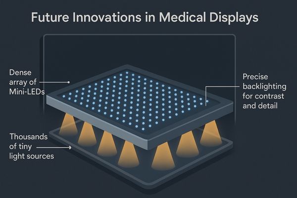
The evolution of detail reproduction is far from over. I see two major innovations on the horizon that will push the boundaries even further. The first is the transition from conventional LED backlights to Mini-LED technology11. Where current local dimming systems have dozens or hundreds of zones, Mini-LED systems will have thousands. This allows for incredibly precise control over the backlight, producing contrast ratios that rival OLED technology but without the associated concerns about lifespan and peak brightness in a clinical setting. The second innovation is the development of smarter, adaptive imaging algorithms. I envision future displays that have AI-powered processors12 built directly into the monitor. This onboard intelligence could analyze the image being displayed—whether it is a CT scan, an X-ray, or an ultrasound—and automatically optimize the rendering parameters to present that specific type of image with maximum clarity. This will make the display a more active and intelligent partner in the diagnostic process. Products like our MD51CHY – 34" 5MP Diagnostic Monitor for X-ray Imaging are built on a platform designed to embrace such future advancements.
Conclusion
The evolution of detail reproduction is a continuous journey. It moves beyond pixels to fuse advanced hardware and intelligent software, enhancing diagnostic confidence and shaping the future of precision medicine. To learn how Reshin is advancing detail reproduction in medical displays, contact us at martin@reshinmonitors.com.
-
Understanding true detail reproduction is crucial for appreciating how modern medical displays enhance image quality and diagnostic accuracy. ↩
-
Exploring AI-driven tools reveals their significant impact on medical imaging, improving efficiency and accuracy in clinical practices. ↩
-
Understanding grayscale linearity is crucial for accurate image representation in medical imaging, ensuring precise diagnostics. ↩
-
Exploring luminance uniformity helps grasp its significance in preventing misinterpretation of medical images, enhancing diagnostic accuracy. ↩
-
Understanding higher bit-depth processing is crucial for appreciating how it enhances image quality in medical diagnostics. ↩
-
Exploring the DICOM standard will provide insights into how medical displays maintain image quality and compliance over time. ↩
-
Explore this link to understand how HDR enhances image quality, especially in critical fields like surgery. ↩
-
Learn about local dimming and its impact on contrast ratios, making visuals more vibrant and detailed. ↩
-
Understanding diagnostic uncertainty can enhance your knowledge of how display quality impacts clinical decisions and patient care. ↩
-
Exploring advancements in mammography can provide insights into how technology improves early detection and patient outcomes. ↩
-
Explore this link to understand how Mini-LED technology enhances display quality and performance, offering superior contrast and brightness. ↩
-
Discover how AI-powered processors can revolutionize medical imaging, optimizing clarity and efficiency in diagnostics. ↩