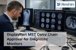
Viewing separate scans creates diagnostic gaps. Clinicians risk missing crucial connections between anatomical and functional data. Medical displays designed for image fusion bridge this gap, ensuring diagnostic confidence.
This article explores how specialized medical displays are essential for multi-modality image fusion. It covers the roles of resolution, color, size, real-time technology, AI integration, and compliance in modern diagnostics and interventions.
Unlocking the full potential of advanced imaging techniques1 like CT, MRI, and PET requires more than just acquiring the data. The true clinical power lies in fusing these modalities together. This fusion creates a single, comprehensive view that overlays functional information onto precise anatomical structures. However, this powerful technique is entirely dependent on the quality of the display. The right medical display2 acts as the critical canvas where these disparate data sets merge into actionable clinical insight. Let’s explore the key display characteristics that make this possible.
High-resolution displays enable accurate fusion of CT, MRI, and ultrasound images
Fusing images on a low-resolution screen causes blurring. This action can misalign anatomical structures and lead to critical errors. A high-resolution display provides the necessary clarity for precise overlays.
High-resolution displays are essential for multi-modality fusion. Their high pixel density prevents data loss and misinterpretation, ensuring that overlapping anatomical and functional details from different scans are aligned with precision.
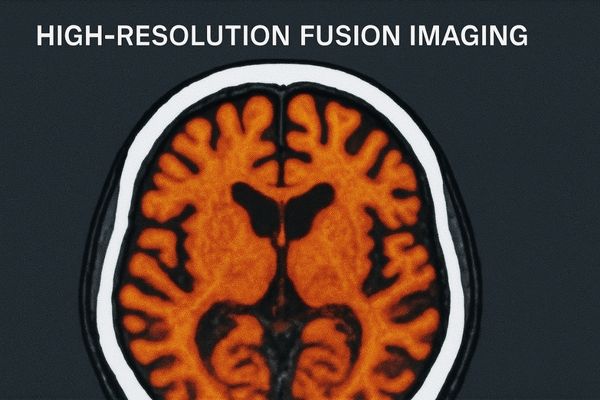
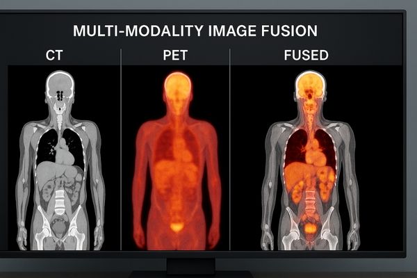
Multi-modality image fusion3 involves digitally overlaying two or more imaging datasets. For example, a high-detail Computed Tomography (CT) scan can be fused with a Magnetic Resonance Imaging (MRI) scan to combine bone structure with soft tissue detail. The success of this process hinges on pixel-level accuracy. If the display lacks sufficient resolution, it cannot render the fine details from both source images simultaneously. This results in aliasing artifacts or blurring, which can make it impossible to determine the exact spatial relationship between structures. A high-resolution medical monitor4, such as our MD120C – 12MP High-Precision Diagnostic Monitor, provides the pixel density required to preserve the integrity of each modality. This ensures that when a radiologist examines a fused image, they can trust that the alignment is accurate, which is fundamental for applications like surgical planning and radiation therapy.
Preserving Data Integrity
The core challenge in image fusion is maintaining the diagnostic value of each original scan. Lower-resolution displays are forced to downsample the image data, effectively discarding pixels to make the image fit. This process can eliminate subtle but clinically significant information.
- CT Scans: Require high spatial resolution to show fine bone fractures or calcifications.
- MRI Scans: Demand high fidelity to differentiate between various soft tissues.
- Ultrasound: Needs pixel accuracy to display real-time blood flow information correctly.
A high-resolution display ensures that the fused composite image is a true and accurate representation of the combined data, not a degraded approximation.
Color accuracy and contrast performance enhance interpretation of multimodal data
Viewing fused functional scans on a standard display washes out vital color data. This weakness makes the functional overlay almost useless. A color-accurate display makes this information clear and quantifiable.
High-end displays are critical when combining functional and anatomical imaging, like PET-CT. Their precise color and contrast are essential for interpreting metabolic activity against anatomical backdrops, guiding complex treatment decisions.
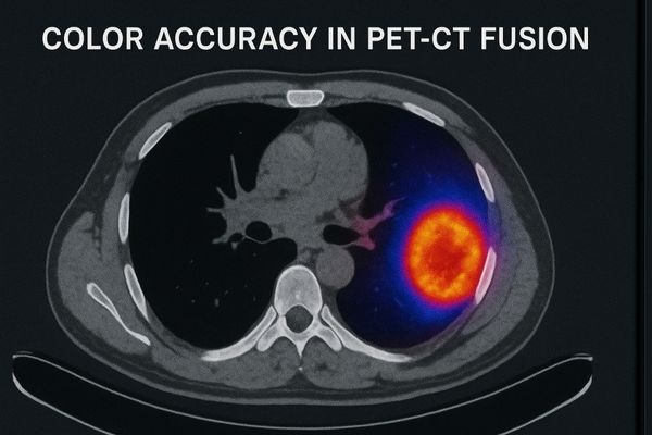
Many fusion techniques involve overlaying functional imaging onto anatomical imaging. A prime example is PET-CT fusion5, where Positron Emission Tomography (PET) data, which shows metabolic activity in color, is laid over a grayscale CT scan. In this context, color is not merely aesthetic; it is quantitative data. The intensity of a color can indicate the severity of a tumor’s metabolic uptake. Therefore, the display must reproduce colors with absolute accuracy and consistency. It must also have a high contrast ratio6 to ensure that subtle color variations from the PET scan are clearly visible against the grayscale background of the CT scan. Our **MD50C – 5MP Color Mammography Monitor** is engineered for exactly this challenge, with a wide color gamut and excellent contrast performance. This capability allows clinicians to confidently assess the extent and activity of diseases, which is vital for oncology staging and treatment response evaluation.
Key Performance Metrics for Fusion Displays
| Feature | Why It Matters for Fusion | Clinical Impact |
|---|---|---|
| Wide Color Gamut | Accurately reproduces the full spectrum of colors used in functional imaging (e.g., PET, SPECT). | Prevents misinterpretation of metabolic activity levels. |
| High Contrast Ratio | Ensures subtle color overlays are clearly visible against detailed grayscale anatomy. | Improves lesion detectability and boundary definition. |
| Luminance Uniformity | Guarantees that the same color value appears identical across the entire screen. | Provides consistent diagnostic information regardless of image position. |
| DICOM Part 14 Calibration | Standardizes grayscale rendering for the anatomical background layer. | Ensures accurate representation of tissue density. |
Large-format and multi-window capabilities support simultaneous viewing of different modalities
Constantly switching between separate monitors is inefficient. This workflow breaks concentration and increases the risk of error. A single, large display showing multiple inputs simplifies comparison and improves focus.
The ability to show multiple modalities side-by-side or in layered fusion modes on a single large screen improves diagnostic and surgical efficiency. It provides a comprehensive view without constant window switching.
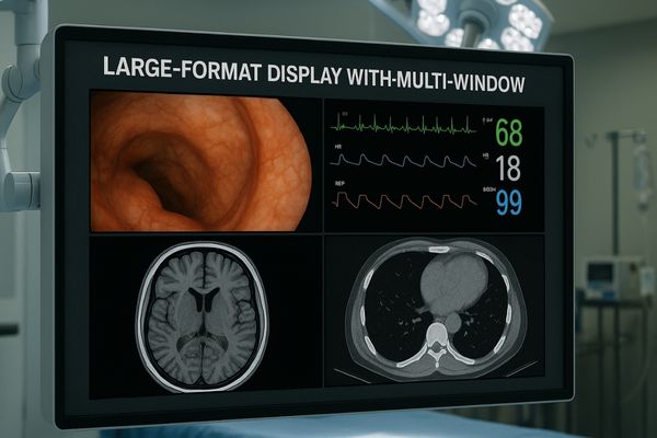
Modern diagnostics and interventions often require the simultaneous review of multiple imaging sources. A radiologist may need to compare a current CT scan with a prior MRI, while a surgeon in a hybrid operating room might need to see live fluoroscopy, endoscopy, and preoperative fused images at the same time. Using separate displays for each modality is cumbersome and cognitively demanding. Large-format displays with multi-window functionality7 solve this problem. Technologies like Picture-by-Picture (PbP)8 allow a single screen to be divided into multiple sections, each displaying a different input source at its native resolution. Our MD46C – Dual-screen Diagnostic Monitor (Single Panel) is specifically designed for this purpose, seamlessly replacing a traditional two-monitor setup. This capability streamlines the clinical workflow, reduces physical clutter, and allows medical professionals to see all relevant information in a single, consolidated view, enhancing decision-making speed and accuracy.
Common Multi-Window Configurations
| Configuration | Description | Best Use Case |
|---|---|---|
| Picture-by-Picture (PbP) | Divides the screen into two or more equal sections for different full-resolution inputs. | Comparing prior and current studies (e.g., CT 2023 vs. CT 2024). |
| Picture-in-Picture (PiP) | Displays a secondary source in a small window overlaid on the primary image. | Monitoring patient vitals while focusing on a primary surgical view. |
| Image Fusion Overlay | A transparent color image (e.g., PET) is layered on top of a grayscale image (e.g., CT). | Integrated analysis in oncology and radiation therapy planning. |
During surgery, outdated static images are insufficient. Surgeons operating on moving organs face immense challenges using old data. Real-time fused imaging provides a live, accurate guide.
Real-time fusion technology on low-latency surgical displays is a game-changer for interventions. It overlays live imaging like ultrasound onto pre-operative scans, providing an accurate, updated map for surgical navigation.
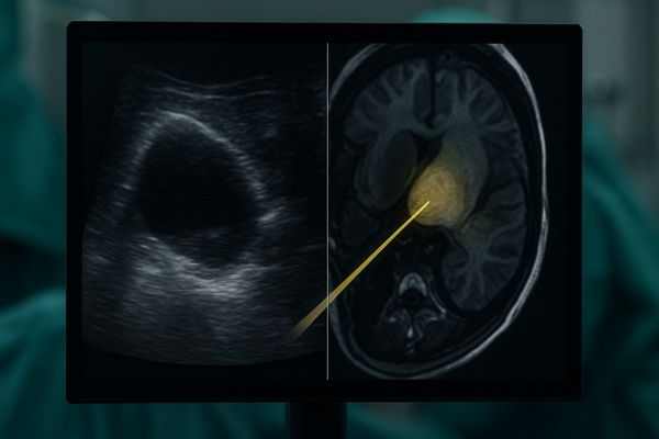
In dynamic environments like the operating room or interventional suite, conditions change rapidly. A pre-operative MRI, for example, shows the state of an organ before an incision is made. During the procedure, tissues can shift. Real-time image fusion addresses this by overlaying live imaging, such as intraoperative ultrasound or fluoroscopy, onto the static pre-operative data. This creates a live, updated roadmap for the surgeon. For this to be effective, the surgical display must have extremely low latency to ensure that what the surgeon sees on the screen perfectly matches what is happening in the patient’s body in real time. Any delay could lead to misguidance. Surgical displays like our MS321PB – 32" 4K Surgical Monitor are built with high-performance video processing to minimize lag, ensuring that surgeons have the immediate visual feedback necessary for precise navigation during complex procedures like tumor resections or catheter placements.
Integration with AI algorithms accelerates detection and diagnosis across modalities
Manually fusing and analyzing multiple image sets is slow and complex. This tedious process increases the cognitive load on clinicians. AI-powered fusion automates this work, providing faster, clearer insights.
AI-enhanced fusion on medical displays accelerates diagnosis. Algorithms can automatically register images and highlight discrepancies or areas of interest, reducing clinician workload and improving the speed and accuracy of detection.
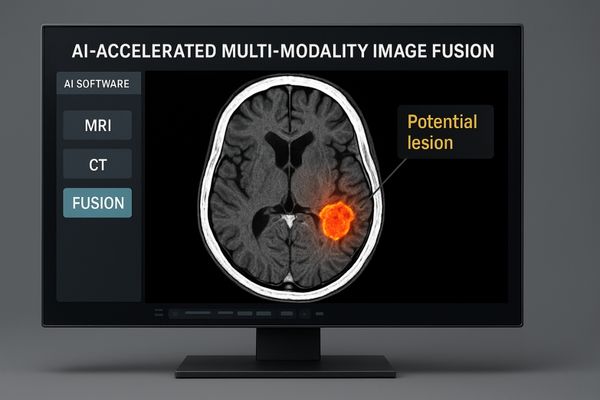
Artificial Intelligence (AI) is transforming multi-modality fusion from a manual process into an automated one. AI algorithms can perform image registration9—the process of perfectly aligning two different scans—in seconds with a level of precision that is difficult to achieve manually. Once the images are fused, other AI tools can analyze the combined data to detect anomalies that might be invisible on a single modality. For instance, an AI might identify a region with high blood flow on an arterial spin labeling (ASL) MRI scan that corresponds to a subtle structural change on a T1-weighted scan, flagging a potential tumor. The diagnostic display serves as the critical interface for this human-AI collaboration. It must clearly render both the fused images and the AI-generated overlays or heatmaps. The MD85CA provides the resolution and clarity needed to visualize these complex, AI-enhanced fused datasets10, empowering radiologists to make faster and more confident diagnoses.
Compliance with standards like DICOM ensures consistent multimodality workflows
If fused images look different on every display, clinical trust is lost. This inconsistency can lead to diagnostic uncertainty and repeated scans. Adherence to standards like DICOM is the only way to guarantee uniformity.
Compliance with standards, especially DICOM for grayscale and color management protocols, is fundamental. It ensures that fused images maintain their clinical accuracy and appear consistent across different systems, departments, and hospitals.
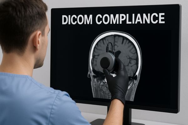
For multi-modality fusion to be a reliable clinical tool, the resulting images must be consistent and reproducible. The DICOM (Digital Imaging and Communications in Medicine)11 standard is the cornerstone of this consistency. Specifically, the DICOM Part 14 Grayscale Standard Display Function (GSDF)12 dictates exactly how a display should render grayscale values. This ensures that a CT or MRI scan will look the same whether it is viewed in the radiology department, a surgeon’s office, or a collaborating hospital. This standardization is even more critical for fused images, as the anatomical grayscale layer must be rendered correctly to provide an accurate backdrop for the functional color overlay. Medical displays like the MD33G – 3MP Grayscale Diagnostic Monitor undergo rigorous factory calibration to ensure strict adherence to the DICOM standard. This embedded compliance guarantees that clinicians are making decisions based on a universally consistent and accurate representation of the image data.
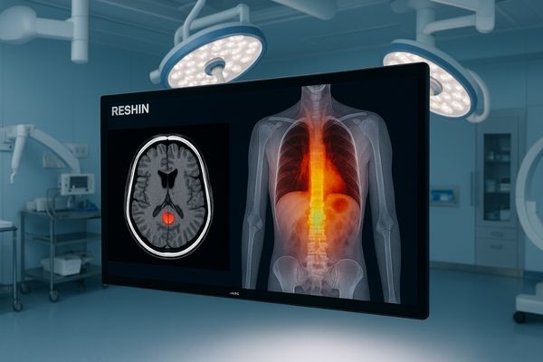
Companies like Reshin deliver specialized solutions tailored for hybrid imaging environments
Generic displays cannot meet the diverse demands of modern fusion imaging. They lack the required brightness, connectivity, and specialized features. This forces compromises in the hybrid OR or diagnostic suite.
I founded this company to solve these specific challenges. Our multimodality-ready displays combine high brightness, superior contrast, and versatile multi-window functionality, making them ideal for advanced hybrid environments.
At Reshin, we understand that multi-modality image fusion13 is not a niche application but a growing standard of care. That is why we engineer our displays to excel in these demanding environments. We focus on delivering the specific combination of features that hybrid imaging requires. This includes high resolution for detail, accurate color for functional data, and high contrast to see both clearly. It also includes versatile connectivity and multi-window processing to manage inputs from CT scanners, C-arms, endoscopes, and other sources simultaneously. Our MS550P – 55" 4K Surgical Monitor, for example, is perfectly suited for the hybrid operating room14. Its large screen size and 4K resolution provide an immersive canvas for viewing complex fused surgical plans, while its high brightness and anti-reflection properties ensure clarity even under bright surgical lights. By focusing on these tailored solutions, we provide medical professionals with the tools they need to leverage the full power of multi-modality fusion.
Conclusion
Specialized medical displays are not optional but essential for multi-modality fusion. They unlock the full potential of advanced imaging, improve diagnostic accuracy, and empower clinicians to make better-informed decisions for superior patient outcomes.
📩 Want to optimize multi-modality imaging with specialized displays? Contact Martin at martin@reshinmonitors.com to explore tailored solutions from Reshin.
-
Understanding advanced imaging techniques can enhance your knowledge of medical diagnostics and their applications. ↩
-
Exploring the importance of medical displays can provide insights into how they impact clinical decision-making and patient care. ↩
-
Explore this link to understand how multi-modality image fusion enhances diagnostic accuracy and treatment planning in medical imaging. ↩
-
Discover why high-resolution medical monitors are crucial for accurate image analysis and how they improve patient outcomes. ↩
-
Understanding PET-CT fusion is crucial for grasping how imaging techniques enhance cancer diagnosis and treatment. ↩
-
Exploring the significance of high contrast ratios can deepen your knowledge of image clarity in medical diagnostics. ↩
-
Explore this link to understand how multi-window functionality enhances medical imaging efficiency and decision-making. ↩
-
Learn about PbP technology to see how it allows simultaneous viewing of multiple imaging sources, improving workflow. ↩
-
Understanding image registration is crucial for grasping how AI enhances medical imaging and diagnostics. ↩
-
Exploring AI-enhanced fused datasets reveals how they improve diagnostic accuracy and efficiency in healthcare. ↩
-
Understanding the DICOM standard is crucial for ensuring consistency in medical imaging, which is vital for accurate diagnoses. ↩
-
Exploring GSDF will enhance your knowledge of how grayscale values are rendered, impacting image interpretation in clinical settings. ↩
-
Explore this link to understand how multi-modality image fusion is revolutionizing healthcare and improving patient outcomes. ↩
-
Discover the advantages of hybrid operating rooms and how they enhance surgical procedures and patient care. ↩