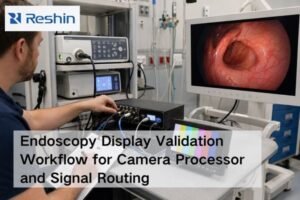
Using standard monitors in clinical settings introduces unacceptable risks. Poor visibility, inaccurate colors, and image lag can lead to diagnostic errors and compromised surgical procedures.
In critical clinical environments such as operating rooms, endoscopy suites, and radiology reading rooms, medical-grade displays address key challenges with features like high brightness, ultra-wide viewing angles, precise DICOM calibration, and low-latency imaging, ultimately safeguarding patient outcomes.
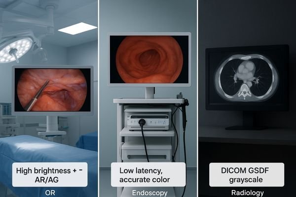
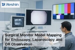
The demands placed on imaging technology vary dramatically across different medical disciplines. An operating room requires displays that can overcome bright ambient light and be seen clearly by an entire team. An endoscopy suite needs to render motion and color with perfect fidelity in real-time. A radiology reading room1 demands absolute consistency in grayscale representation to reveal the most subtle pathologies. Consumer-grade monitors are not designed to meet any of these specialized, high-stakes requirements. This article explores three key clinical environments—the operating room, the endoscopy suite, and the radiology department—to demonstrate how purpose-built medical-grade displays2 solve their unique and fundamental imaging challenges.
Operating Room (OR): High Brightness & Anti-Glare
Intense surgical lights wash out standard monitor screens. This glare and low contrast can hide critical anatomical details, jeopardizing the safety of the procedure.
Surgical monitors combat bright OR lighting with high luminance (over 500 cd/m²) and anti-reflective, optically bonded panels. This ensures a clear, high-contrast view by preventing glare and reflections from obscuring the image.
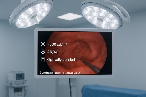
The operating room is one of the most challenging environments for visual displays due to extremely high levels of ambient light from powerful overhead surgical lamps. A standard monitor would be rendered useless, its image appearing faded and washed out. Medical-grade surgical displays are specifically engineered to overcome this challenge. They feature exceptionally high-brightness backlights, often exceeding 500 cd/m² and sometimes reaching over 800 cd/m². This high luminance ensures the image has enough power to maintain its contrast and color saturation. This is complemented by advanced anti-reflective (AR) protective glass, which is often optically bonded directly to the LCD panel. This bonding process eliminates the internal air gap that causes reflections and significantly improves contrast. A 4K surgical monitor3 like the MS275PA combines these technologies to deliver a sharp, vibrant, and glare-free image, allowing the surgical team to see every detail with clarity, regardless of the intense lighting conditions.
Operating Room (OR): Ultra-Wide Viewing Angles
When surgical team members view a standard monitor from the side, the image distorts. This inconsistency means not everyone sees the same vital information, risking miscommunication.
Medical-grade displays provide ultra-wide 178° viewing angles, ensuring every team member sees the same high-fidelity image. This consistency is crucial for effective collaboration, communication, and safety during surgical procedures.
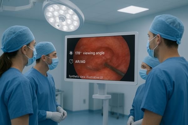
Surgery is a collaborative effort, with surgeons, assistants, nurses, and anesthesiologists all needing to see the same visual information simultaneously. On a monitor with a limited viewing angle, a person viewing from the side would see a dimmer, color-shifted image compared to someone viewing it head-on. This discrepancy is dangerous, as it can lead to misinterpretation and flawed communication. To solve this, surgical displays are built with panel technologies, primarily In-Plane Switching (IPS)4, that provide ultra-wide, symmetric viewing angles of 178° both horizontally and vertically. This guarantees that the image’s brightness, contrast, and color fidelity remain consistent and accurate from virtually any position in the OR. Whether a team member is standing beside the lead surgeon or across the room, they can trust that they are seeing the exact same clinical picture. A large-format display like the 55-inch MS550P is ideal for ensuring this shared, unified view in complex procedures involving large teams.
Operating Room (OR): Multi-Input Connectivity & Low Latency
A delay between a surgeon’s action and the image appearing on screen is unacceptable. This input lag can lead to imprecise movements and compromise procedural safety.
Surgical displays feature versatile high-bandwidth inputs like HDMI, DisplayPort, and 3G/12G-SDI. Their low-latency processing ensures real-time video from various OR devices is displayed smoothly, enabling precise control during minimally invasive surgery.
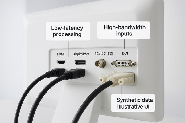
A modern operating room is a hub of advanced imaging equipment, including endoscopes, laparoscopes, C-arms, and surgical cameras. A surgical display must be able to connect to these diverse sources and display their video feeds with virtually zero delay. Medical monitors are equipped with a comprehensive range of high-bandwidth inputs, including HDMI, DisplayPort, and critically, 3G-SDI or 12G-SDI5 (Serial Digital Interface), which is the standard for transmitting uncompressed, high-fidelity video over long distances in professional environments. Beyond connectivity, the monitor’s internal processing is optimized for extremely low latency. This ensures that the live video appears on screen in real time, perfectly synchronized with the surgeon’s movements. During a delicate minimally invasive procedure, this instantaneous feedback is essential for precise instrument control and safety. A versatile monitor such as the MS430PC provides the necessary connectivity and low-latency performance6 to serve as a reliable central imaging hub in the OR.
Operating Room (OR): Optical Bonding & Sterility
Standard monitors have seams and gaps where fluids and contaminants can collect. This makes proper disinfection impossible and poses a significant risk in a sterile environment.
Surgical displays feature optically bonded, fully sealed front panels that prevent dust and fluid ingress. Their flat, seamless surfaces and rugged design allow for easy, effective cleaning, meeting the strict hygiene requirements of the OR.

Hygiene and sterility are non-negotiable in the operating room. Medical equipment must be designed to withstand rigorous cleaning and disinfection protocols without degrading. Consumer monitors, with their plastic bezels, seams, and vents, are impossible to properly sterilize. Surgical displays solve this challenge with purpose-built designs. They often feature a single, edge-to-edge sheet of protective glass that is optically bonded to the LCD panel. This creates a fully sealed, IP-rated front surface that is impervious to dust, splashes, and cleaning agents. The housing is typically smooth and seamless, eliminating crevices where contaminants could accumulate. This robust, easy-to-clean construction ensures the display can be quickly and thoroughly disinfected between procedures, upholding the integrity of the sterile field. Models like the MS321PC are engineered with these hygienic design principles7, making them a safe and durable choice for the demanding OR environment.
Endoscopy: Real-Time HD Imaging
Flickering or blurry video during an endoscopic procedure is disorienting. It can cause eye fatigue and make it difficult to navigate delicate internal structures quickly.
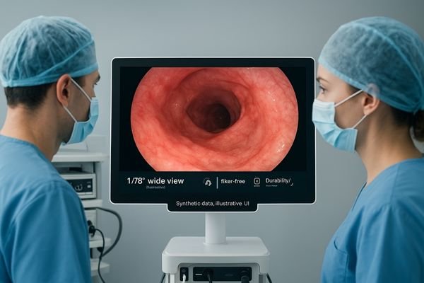
Endoscopy displays deliver Full HD or 4K resolution with fast response times and flicker-free backlighting. This ensures smooth, clear, and immediate visualization of rapid endoscopic movements, reducing eye strain and improving procedural control.
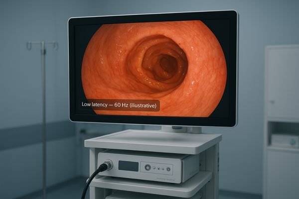
During an endoscopic examination, the clinician relies on a live video feed to navigate complex anatomical pathways. The display must render this motion smoothly and with perfect clarity. Any lag, motion blur, or flicker can cause disorientation, lead to eye fatigue during long procedures, and potentially cause the physician to miss a subtle finding. Endoscopy monitors are optimized for real-time performance. They feature high-resolution panels8, typically Full HD or 4K, to show fine detail. More importantly, their electronics are engineered for fast pixel response times and are paired with flicker-free backlighting technologies. This combination eliminates distracting visual artifacts and ensures that the on-screen image keeps pace with even the most rapid movements of the endoscope tip. A dedicated endoscopy monitor like the MS247SA provides the stable, crystal-clear imaging necessary for confident navigation and diagnosis, allowing the physician to focus entirely on the patient.
Endoscopy: Accurate Color & Gamma Fidelity
If a display shows inaccurate colors, a physician might misinterpret healthy tissue as inflamed. This could lead to unnecessary biopsies or missed diagnoses of subtle pathologies.
Endoscopy monitors use high-bit gamma processing and adhere to color standards like BT.709. This true-to-life color and gamma fidelity allows clinicians to accurately distinguish between subtle variations in mucosal and vascular tissue.
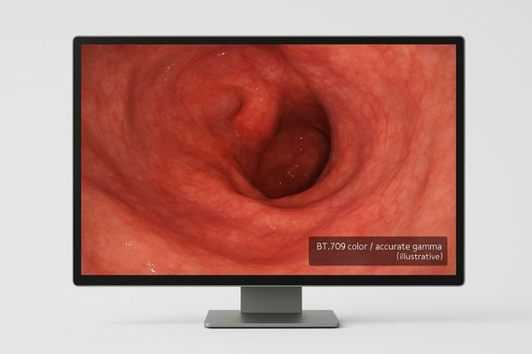
In endoscopy, color is not merely aesthetic; it is a primary diagnostic tool. The ability to distinguish between subtle shades of red, pink, and white is critical for identifying inflammation, dysplasia, and vascular abnormalities. Consumer monitors are not calibrated for clinical accuracy and can render these vital colors incorrectly. Medical endoscopy monitors solve this by using advanced color science9. They employ high-bit-depth gamma processing10 and are calibrated to adhere to international color standards, such as BT.709 for HD video. This ensures that the colors captured by the endoscope’s camera are reproduced on the screen with exceptional fidelity. The precise gamma curves allow for the visualization of subtle details in both bright and shadowed areas of the image. Displays like the MS321PB provide this level of color and gamma accuracy, giving clinicians confidence that they are seeing a true-to-life representation of the patient’s internal anatomy.
Endoscopy: Extensive Connectivity
An endoscopy suite uses scopes and processors from different eras and manufacturers. A new display that cannot connect to older analog equipment disrupts workflow and limits options.
Medical endoscopy monitors provide comprehensive connectivity, including modern digital (SDI, DVI) and legacy analog (S-Video, Composite) inputs. This guarantees plug-and-play compatibility with a wide range of new and existing endoscopic systems.
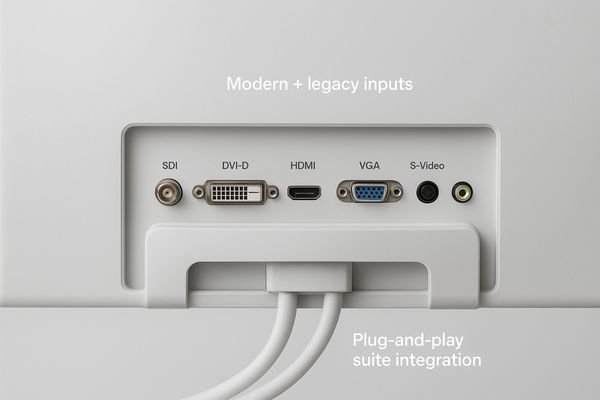
An endoscopy suite is often an ecosystem of equipment acquired over many years from various manufacturers. It can include the latest 4K digital video processors alongside older, standard-definition systems that are still in service. A display must be able to connect to all of them seamlessly. While a standard monitor may only have HDMI and DisplayPort, a dedicated endoscopy display11 provides a much more extensive range of I/O options. This includes modern digital inputs like SDI and DVI, but also crucial legacy analog inputs such as S-Video, Composite, and VGA. This "universal" connectivity ensures that the monitor can be integrated into any endoscopy tower, regardless of its age or manufacturer, without requiring cumbersome and signal-degrading adapters. A monitor like the MS192SA is designed with this comprehensive I/O support, offering the flexibility and plug-and-play convenience that a busy clinical environment demands.
Endoscopy: Wide Viewing Angle & Reliability
During a procedure, nurses and assistants need to see the screen clearly from the side. On a standard monitor, the image would be distorted, hindering their ability to assist effectively.
High-grade endoscopy monitors feature wide 178° viewing angles for team-wide visibility. Their robust construction and high MTBF ratings ensure reliable, continuous operation and reduce eye fatigue during long, demanding procedures.
Like the OR, the endoscopy suite is a collaborative space. It is essential that everyone on the team, not just the primary physician, can see a clear image. Endoscopy monitors utilize IPS or similar panel technologies to deliver the same 178° wide viewing angles found in surgical displays, ensuring consistent image quality for all viewers. Beyond visibility, reliability is paramount. These procedures can be long, and the equipment is often in continuous use throughout the day. Endoscopy displays12 are built for this reality. They feature rugged construction and are subjected to rigorous testing to achieve high Mean Time Between Failures (MTBF)13 ratings, often exceeding 30,000 hours. Flicker-free screen technology also helps to reduce operator eye fatigue over the course of a long day of procedures. A reliable, ergonomic display like the MS270P provides the durability and consistent performance needed for the demanding endoscopy environment.
Radiology: Precise DICOM Calibration
Viewing a chest X-ray on an uncalibrated monitor is dangerous. A faint, low-contrast nodule that is clearly visible on a DICOM-calibrated display could be completely invisible.
Radiology displays are factory-calibrated to the DICOM Part 14 standard. This ensures standardized grayscale reproduction, making image interpretation consistent and enabling radiologists to perceive subtle abnormalities that would be missed on consumer screens.
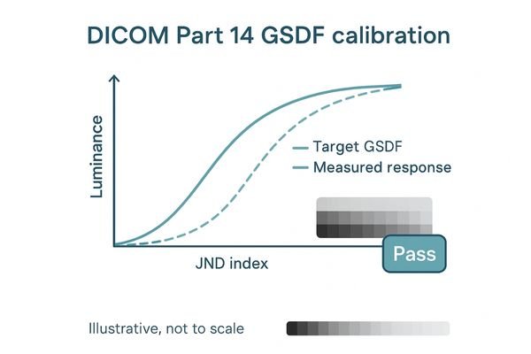
In radiology, diagnosis hinges on the consistent and accurate interpretation of grayscale images like X-rays, CT scans, and MRIs. The human eye’s perception of brightness is not linear, and the DICOM Part 14 Grayscale Standard Display Function (GSDF)14 was created to standardize monitor luminance to match this perception. Medical diagnostic displays are meticulously calibrated at the factory to conform to this standard. This ensures that a specific pixel value will correspond to the same perceived brightness on any DICOM-compliant monitor, anywhere in the world. This consistency is crucial for detecting subtle, low-contrast pathologies like faint nodules or fine fractures. Unlike consumer monitors, which have no such standard, diagnostic displays use high-bit internal look-up tables (LUTs)15 and often have built-in sensors for automatic quality control to maintain this calibration over time. Displays like the MD45C provide this essential DICOM compliance, forming the foundation of accurate radiological diagnosis.
Radiology: High Contrast & Resolution
Trying to diagnose from a CT scan on a low-resolution office monitor is inefficient and risky. Fine details are lost, forcing constant zooming and increasing the chance of oversight.
Radiology monitors offer high native contrast ratios and resolutions from 2MP up to 12MP. This allows radiologists to visualize fine anatomical structures and low-contrast lesions with exceptional clarity, improving both diagnostic accuracy and efficiency.
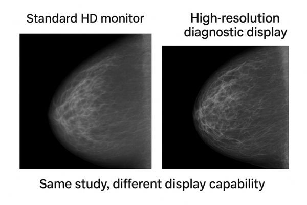
Radiological images are incredibly data-rich. To see all the detail captured by a modern CT or MRI scanner, the display must have sufficient resolution and contrast. Diagnostic monitors offer resolutions far beyond standard HD, ranging from 2MP or 3MP for general radiology up to 5MP for mammography, and even higher for multi-modality studies. A higher resolution means more pixels are available to display the image, revealing finer details without requiring constant panning and zooming. This is complemented by a high native contrast ratio16 (typically 1000:1 or greater), which enhances the visibility of low-contrast abnormalities against their background tissue. For example, a flagship 12MP display17 like the MD120C can show multiple images from different modalities side-by-side in their full native resolution, providing an unparalleled overview for complex cases. This combination of high resolution and high contrast is fundamental to confident and efficient diagnostic interpretation.
| Display Type | Typical Resolution | Common Clinical Application |
|---|---|---|
| Clinical Review | 1-2 MP | Reviewing reports, secondary images |
| General Radiology | 2-3 MP | CT, MRI, X-ray interpretation |
| Mammography | 5 MP | Screening and diagnostic mammograms |
| Multi-Modality | 6-12 MP | Fused imaging (PET/CT), complex cases |
Radiology: Multi-Mode Display & Workflow
Switching between different windows for image review and patient reports is slow. This constant task-switching breaks concentration and contributes to radiologist fatigue and burnout.
Many diagnostic displays support picture-by-picture or other multi-mode layouts. This allows radiologists to view grayscale diagnostic images alongside color reference images or reports on a single screen, streamlining workflow and reducing physical strain.
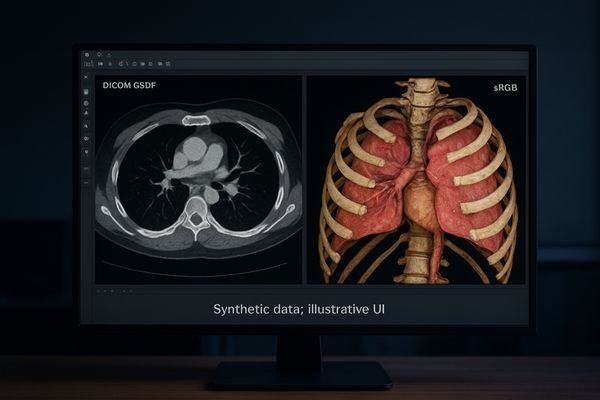
A radiologist’s workflow often involves comparing current studies to priors or reviewing images alongside the corresponding clinical report. Constantly switching between different monitors or applications is inefficient and ergonomically taxing. To solve this, advanced diagnostic displays incorporate workflow enhancement features18. Many support picture-by-picture (PBP) or picture-in-picture (PIP) modes, allowing the screen to be partitioned to display inputs from two different sources simultaneously. Some can even apply different calibrations to different parts of the screen, such as showing a DICOM-calibrated grayscale image next to a color image in sRGB mode. This allows the radiologist to have all the necessary information consolidated on a single, unified desktop. A versatile monitor like the MD85CA offers these multi-mode capabilities19, which help to streamline the reading process, reduce head and eye movement, and ultimately improve diagnostic efficiency and focus.
Radiology: Stable Brightness & Uniformity
All monitors dim over time. An aging, unmanaged display can fall out of DICOM compliance, leading to inconsistent image quality and potentially missed diagnoses.
Diagnostic displays use Continuous Brightness Stabilization and uniformity compensation technologies. These systems counteract backlight aging and ensure consistent, even brightness across the entire screen, maintaining DICOM compliance and diagnostic quality over the long term.
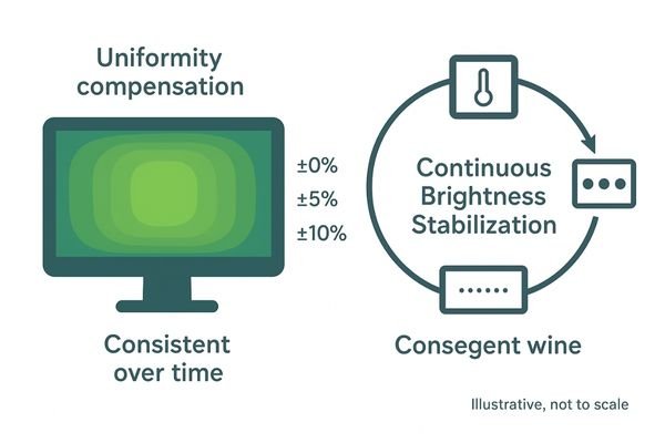
A diagnostic display must not only be accurate on its first day of use, but for thousands of hours thereafter. The backlights in all LCDs inevitably dim with age, which can cause the monitor to fall out of DICOM compliance. Medical displays are engineered for long-term stability to counteract this effect. They incorporate Continuous Brightness Stabilization20 (CBS) systems, which use a front or backlight sensor to constantly measure the luminance and adjust the power to maintain a consistent output. Furthermore, they feature uniformity compensation technology21. This corrects for the slight variations in brightness that can occur from the center to the corners of any LCD panel, ensuring a perfectly even image. These stabilization and uniformity systems, found in high-performance monitors like the MD32C, work together to guarantee that the image quality remains stable and reliable throughout the monitor’s entire lifespan, protecting the integrity of the diagnostic process.
Conclusion
Medical-grade displays solve critical challenges by delivering specialized, reliable performance in surgery, endoscopy, and radiology, directly enhancing diagnostic confidence, procedural safety, and overall patient care.
👉 To explore the right medical display solutions for your clinical applications, connect with Martin at martin@reshinmonitors.com — we’ll help match the best technology to your needs.
-
Learn about the critical standards for imaging technology in radiology to ensure accurate diagnosis and patient care. ↩
-
Explore how medical-grade displays enhance imaging quality and meet the specific needs of various medical environments. ↩
-
Discover the benefits of 4K surgical monitors for improved image clarity and detail during surgical procedures. ↩
-
Explore this link to understand how IPS technology enhances viewing angles and image quality in surgical settings ↩
-
Understanding 3G-SDI and 12G-SDI is crucial for anyone involved in medical imaging, ensuring high-quality video transmission. ↩
-
Exploring low-latency performance will highlight its significance in enhancing surgical precision and patient safety. ↩
-
Learn about the importance of hygienic design principles in medical equipment to maintain sterility and prevent infections. ↩
-
Learn about the significance of high-resolution panels in medical displays for enhanced detail and accuracy in diagnostics. ↩
-
Understanding color science is crucial for accurate medical imaging, ensuring precise diagnosis and treatment. ↩
-
Exploring high-bit-depth gamma processing reveals how it enhances image quality, vital for effective medical evaluations. ↩
-
Explore this link to understand how specialized endoscopy displays enhance connectivity and performance in clinical environments. ↩
-
Explore this link to discover top-rated endoscopy displays that ensure reliability and image quality in surgical settings. ↩
-
Understanding MTBF is crucial for evaluating the reliability of medical equipment; this link provides valuable insights. ↩
-
Understanding GSDF is vital for ensuring accurate image interpretation in radiology, enhancing diagnostic precision. ↩
-
Exploring LUTs will reveal how they improve image quality and consistency in medical diagnostics, crucial for accurate assessments. ↩
-
Understanding high native contrast ratios is crucial for evaluating image quality in diagnostic monitors, enhancing diagnostic accuracy. ↩
-
Exploring the advantages of a 12MP display can reveal how it improves diagnostic capabilities and image clarity in medical imaging. ↩
-
Explore this link to understand how workflow enhancement features can significantly improve efficiency in radiology. ↩
-
Discover how multi-mode capabilities in monitors can streamline the diagnostic process and enhance radiologist productivity. ↩
-
Understanding CBS can help you appreciate how medical displays maintain image quality over time, ensuring accurate diagnostics. ↩
-
Exploring this technology reveals how it enhances image consistency, crucial for reliable medical diagnostics. ↩