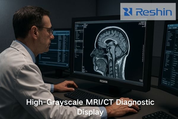
Radiologists reviewing MRI and CT scans frequently miss subtle pathology due to insufficient grayscale rendering on substandard displays. Could your diagnostic accuracy be compromised by outdated or inadequate medical monitors?
Choosing the right high-grayscale medical diagnostic display is essential for MRI and CT accuracy in 2025. The following three models stand out as top performers:
- MD33G — High-Grayscale Diagnostic Medical Display
- MD46C — High-Brightness Diagnostic Medical Display
- MD32C — High-Grayscale Standard Diagnostic Display
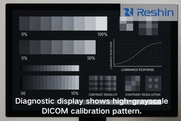
These displays deliver DICOM Part 14 compliance, stable luminance performance, strong uniformity control, and deep grayscale rendering—ensuring consistent visualization of subtle MRI/CT tissue variations and potential pathologies.
As an engineer specialized in medical display calibration and integration, I’ve seen firsthand how display quality directly affects diagnostic confidence. Across my deployments in radiology departments, the right high-grayscale diagnostic monitor consistently improves visibility of subtle MRI/CT pathologies and streamlines the clinical workflow. This article summarizes the essential features of modern high-grayscale displays and reviews the top three models delivering reliable performance in 2025.
Key Features of a High-Grayscale Medical Diagnostic Display for MRI and CT in 2025
What separates a true diagnostic-quality display from standard medical monitors when evaluating subtle grayscale differences in MRI and CT imaging?
Across my workstation deployments, the most critical requirement for MRI and CT displays is long-term stability in grayscale depth and luminance performance. MRI and CT rely on subtle grayscale transitions to visualize vascular structures, soft-tissue gradients, and organ boundaries. A high-quality diagnostic monitor must deliver DICOM Part 14 accuracy, stable luminance, strong uniformity correction, and deep grayscale rendering—especially for low-contrast pathologies.
Essential Technical Requirements
When evaluating diagnostic monitors for MRI and CT applications, several technical capabilities stand out:
-
Deep Grayscale Rendering Capability
- Minimum 10-bit grayscale (1024 gray levels)
- Preferably 12-bit LUT for smoother transitions
- Ability to follow the DICOM GSDF curve without banding
-
Long-Term Luminance Stability
- Integrated luminance stabilization
- Automatic brightness feedback loop
- Drift kept within clinically acceptable limits with minimal recalibration
-
Panel Uniformity Compensation
- Factory and hardware-assisted correction
- Variation kept within medical standards
- Ensures consistent grayscale across the entire panel
-
Strict DICOM Part 14 Compliance1
- Hardware-level calibration preferred
- Automated conformance verification
- Consistent grayscale rendering across sessions
-
Multi-modality Flexibility
- Dedicated modes for MRI, CT, DR, ultrasound
- Consistent gamma behavior
- Support for hybrid grayscale/color workflows
All three displays featured in this article meet or exceed these grayscale and luminance performance requirements for MRI and CT reading.
Workflow-Enhancing Features
| Feature | Clinical Benefit | Impact on Diagnostic Accuracy |
|---|---|---|
| Front sensor QA | Automated compliance without interruptions | Ensures consistent calibration |
| Multi-input capability | Enables comparison of current/prior studies | Helps detect subtle progression |
| Integrated calibration software | Simplified QA | Maintains stable grayscale rendering |
| Remote management | Centralized monitoring | Reduces risk from calibration drift |
| Ergonomic design | Less fatigue in long reading sessions | Supports consistent visual acuity |
MD33G and MD46C implement nearly all of these workflow-enhancing features, while MD32C supports them at a more cost-efficient level.
Top 3 High-Grayscale Medical Monitors for MRI & CT in 2025
MD33G High-Grayscale Diagnostic Medical Display
The MD33G is my preferred choice for MRI/CT primary diagnosis rooms. Its high brightness, excellent grayscale differentiation, and robust uniformity correction make it ideal for visualizing low-contrast structures and density variations. Hardware DICOM calibration and long-term luminance stabilization preserve consistent grayscale performance throughout long reading sessions.
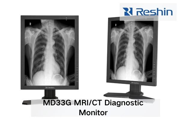
The MD33G serves as a premium option for MRI and CT interpretation. Its 3MP resolution and high-accuracy grayscale rendering make it suitable for demanding clinical environments.
Technical Specifications
| Specification | Value | Clinical Significance |
|---|---|---|
| Resolution | 3MP (2048×1536) | Standard for MRI/CT protocols |
| Maximum Brightness | 1000 cd/m² | Strong visibility in low-contrast regions |
| Contrast Ratio | 1500:1 | Clear differentiation of subtle densities |
| Bit Depth | 10-bit native | Smooth grayscale without banding |
| Calibration2 | Hardware DICOM + front sensor | Reliable conformance with minimal manual effort |
| Luminance Stabilization | CBS system | Stable brightness over years of usage |
| Viewing Angle | 178°/178° | Ideal for collaborative review |
| Uniformity | Advanced hardware correction | Consistent grayscale across the panel |
Optimal Clinical Applications
- Low-contrast CT lesion evaluation — Enhances visibility of subtle hepatic or pulmonary nodules
- MRI T1/T2 subtle structure analysis — Improves signal differentiation
- Multi-slice CT review — Ensures consistent soft-tissue contrast across sequential slices
- Thin-slice reconstructions — Maintains grayscale fidelity under high-detail settings
MD33G consistently demonstrates strong performance in detecting subtle MRI/CT pathologies across my deployments.
MD46C High-Brightness Diagnostic Medical Display
The MD46C offers high brightness and deep grayscale capability, making it suitable for multi-modality reading where radiologists compare MRI, CT, and DR images. Its wide viewing angle and strong luminance retention support both individual and collaborative diagnostic review.
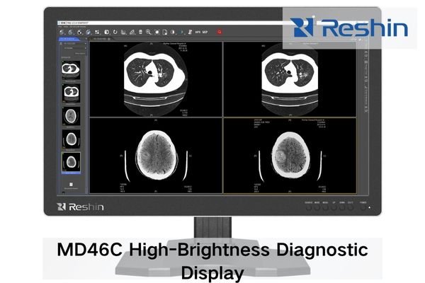
The MD46C is ideal for high-volume radiology departments requiring efficient comparative workflows.
Technical Specifications
| Specification | Value | Clinical Significance |
|---|---|---|
| Resolution | 2 × 4MP (2048×2560 per screen) | Excellent for side-by-side comparisons |
| Maximum Brightness3 | 800 cd/m² | Suitable for varied room lighting |
| Contrast Ratio | 1400:1 | Strong tissue differentiation |
| Bit Depth | 10-bit with 12-bit LUT | Smooth grayscale transitions |
| Calibration | Automated DICOM verification | Maintains GSDF conformance |
| Luminance Stabilization | Integrated CBS | Stable brightness output |
| Viewing Angle | 178°/178° | Supports group interpretation |
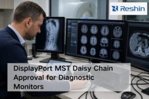
| Inputs | DP, HDMI, DVI | Flexible modality compatibility |
Optimal Clinical Applications
- Comparative reading workflows — Current vs. prior MRI/CT
- Multi-modality correlation — MRI + CT + DR
- High-volume reading — Efficient layout for many open studies
- Teaching environments — Wide viewing angles aid group learning
- Hybrid clinical workflows — Radiology + reporting + visualization
For community hospitals and regional imaging centers, MD46C offers premium efficiency without the cost of ultra-high-resolution hardware.
MD32C High-Grayscale Diagnostic Display
The MD32C is a well-balanced high-grayscale display suited for standard radiology rooms. With strong uniformity, reliable grayscale performance, and low maintenance cost, it is widely adopted in budget-conscious environments seeking consistent MRI/CT diagnostic capability.

The MD32C delivers dependable essential performance for daily radiology reading tasks.
Technical Specifications
| Specification | Value | Clinical Significance |
|---|---|---|
| Resolution | 3MP (2048×1536) | Suitable for routine MRI/CT interpretation |
| Maximum Brightness | 700 cd/m² | Adequate for standard reading rooms |
| Contrast Ratio | 1200:1 | Good tissue differentiation |
| Bit Depth | 10-bit | Smooth grayscale |
| Calibration | Software-based DICOM | Consistent with regular QC checks |
| Luminance Stabilization | Basic stabilizer | Reduces calibration frequency |
| Uniformity | Digital correction | Improved screen consistency |
| Power | 55W | Low cost of ownership |
Optimal Clinical Applications
- Standard MRI/CT reading rooms
- Secondary review stations
- Training environments
- Budget-constrained radiology centers
- Regional and community hospitals needing reliable diagnostic visualization4
For institutions upgrading from non-medical monitors, MD32C represents a strong entry-level diagnostic solution.
Why Radiology Departments Must Choose the Right High-Grayscale Diagnostic Display
MRI and CT interpretation relies heavily on precise grayscale rendering. Inadequate displays can obscure low-contrast lesions and compromise evaluation accuracy. Across my deployments, I’ve seen brightness decay, calibration drift, and poor uniformity directly lead to longer reading times and re-reads. Evaluating resolution, luminance stability, uniformity, and PACS integration is essential when selecting diagnostic monitors.
Clinical Impact of Display Quality
1. Diagnostic Accuracy
Suboptimal displays may cause:
- Loss of low-contrast details
- Inconsistent grayscale across screen regions
- Perceptual shifts during long sessions
- GSDF deviations affecting modality consistency
2. Reading Efficiency5
High-quality displays improve:
- Interpretation speed
- Visual comfort
- Workflow continuity
- Multi-modality transitions
3. Long-Term Reliability
| Factor | Poor Stability | High-Quality Display Benefit |
|---|---|---|
| Luminance decay | Gradual loss of visibility | Consistent diagnostic clarity |
| Calibration drift | Frequent QC failures | Reduced maintenance workload |
| Panel aging | Non-uniform artifacts | Preserved uniformity |
| Hardware reliability | More downtime | Stable department operations |
A suitable high-grayscale monitor reduces workflow friction and supports consistent MRI/CT interpretation over many years.
Real-world Case Study: High-Grayscale Medical Displays in an MRI/CT Diagnostic Environment
In a European hospital upgrade, radiologists struggled to detect low-density CT lesions due to aging displays with grayscale inconsistency. We installed MD33G as the primary diagnostic display and MD46C for comparative reading. The updated workstation improved soft-tissue differentiation and reduced QA workload. Reading efficiency improved noticeably, and re-reads decreased.
Challenge Identification
Clinical challenges:
- Difficulty detecting subtle CT lesions
- Inconsistent grayscale across workstations
- Excessive MRI window/level adjustments
Technical issues:
- Significant luminance drift
- GSDF deviations
- Non-uniform panels
Workflow impact:
- Longer reading times
- More consultation requests
- Frequent QC failures
Solution Implementation
-
Primary Stations
- MD33G with hardware DICOM calibration
- Integrated into PACS with consistent display profiles
-
Comparative Stations
- MD46C dual-screen configuration
- Calibrated screen-pair matching
-
Quality Assurance
- Daily automated checks
- Monthly calibration verification
Measured Outcomes
Clinical:
- Notable improvement in detecting subtle CT lesions
- Better MRI tissue characterization
- More consistent reporting
Workflow:
- Faster reading times for complex studies
- Fewer technical interventions
- Seamless workstation consistency
Long-term:
- Lower display maintenance cost
- Extended refresh cycle
- Reduced radiologist fatigue
These results reflect one institution’s experience; actual performance may vary based on workflow and infrastructure.
Why Choose Reshin as Your Radiology Display Partner?
In radiology deployments, I prioritize luminance stability, lifecycle management, flexibility, and strong service support. Reshin’s high-grayscale diagnostic displays deliver CBS stabilization, hardware uniformity correction, multi-signal support, and long-term calibration strategies—enabling consistent MRI/CT diagnostic environments. With OEM/ODM capabilities, rapid spare-parts support, and professional engineering teams, Reshin provides reliable long-term partnership value.
Technical Excellence
- Advanced Stabilization (CBS) — Predictive, real-time, and aging-aware
- Comprehensive Calibration Ecosystem — Automated verification + remote management
- Engineering-Driven Design — Developed with radiologist workflow insights
Partnership Advantages
| Element | Reshin Advantage | Customer Benefit |
|---|---|---|
| Lifecycle Support | Minimum 7-year product continuity | Reliable long-term planning |
| Implementation | Dedicated specialists | Optimized configurations |
| Technical Support | Direct engineering access | Fast issue resolution |
| OEM/ODM | Customizable | Tailored clinical solutions |
| QA & Compliance | ISO 13485 | Reliable manufacturing quality |
Global Support Infrastructure
- Regional technical centers
- Continuous education programs
- Long-term partnership approach
This support ecosystem ensures sustained clinical performance and maximum ROI for radiology departments.
Conclusion
As MRI and CT diagnostic demands continue to rise in 2025, selecting a reliable high-grayscale medical diagnostic display is essential for maintaining clinical accuracy and workflow efficiency. The MD33G, MD46C, and MD32C each support different radiology scenarios—from advanced primary diagnosis to cost-effective daily reading—providing stable luminance, consistent uniformity, and dependable grayscale performance.
From my engineering experience, choosing the right display directly improves subtle lesion visibility and reduces unnecessary workflow interruptions. If your radiology department is planning a workstation upgrade or evaluating MRI/CT diagnostic display options, feel free to contact our engineering team for a tailored recommendation.
📧 info@reshinmonitors.com
🌐 https://reshinmonitors.com/
-
Exploring this compliance helps ensure that monitors meet essential standards for medical imaging accuracy. ↩
-
Exploring calibration methods can reveal how to achieve reliable conformance and improve image quality in clinical settings. ↩
-
Understanding maximum brightness helps ensure optimal visibility in various lighting conditions, crucial for accurate diagnostics. ↩
-
This resource will provide insights into techniques and technologies that enhance diagnostic visualization, crucial for accurate patient care. ↩
-
Exploring Reading Efficiency can enhance your knowledge on optimizing workflows and improving diagnostic processes in healthcare. ↩