Choosing monitors for a cardiac catheterization lab presents many challenges. Incorrect displays can hinder critical procedures. Understanding specific features ensures optimal performance and patient safety in this demanding environment.
Monitors suited for cardiac catheterization labs must offer high resolution, precise grayscale accuracy, low latency, and robust construction. They need to support real-time imaging like fluoroscopy and angiography, and handle the cath lab’s unique environmental demands.
The cardiac catheterization lab, or cath lab, is a specialized environment where complex, image-guided procedures are performed. The visual information presented to the cardiology team is paramount for accurate diagnosis and intervention. Therefore, the choice of medical displays is not a trivial one. In the following sections, I will explore the key characteristics that define a suitable monitor for this critical care area, covering everything from imaging requirements to physical integration.
What are the key imaging requirements in a cath lab environment?
Cath labs operate at a fast pace, with critical decisions made in real-time. Displayed images can be complex and varied. It is vital to know what specific imaging capabilities are essential for success.
Key imaging requirements in a cath lab include real-time display of fluoroscopy and angiography, often alongside physiological data. Monitors must support multi-window layouts, high frame rates for smooth motion, and very low latency for immediate visual feedback.
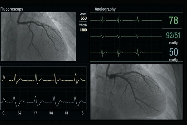
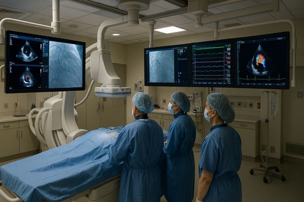
The imaging needs within a cardiac catheterization lab are multifaceted and demanding. Primarily, monitors must clearly display dynamic imaging modalities. Real-time fluoroscopy, which provides live X-ray video, is fundamental for guiding catheters and devices through blood vessels. Angiography, involving the injection of contrast media to visualize vessel lumens, requires high clarity to detect stenoses or anomalies. These live video feeds necessitate high frame rates, typically 60 frames per second or higher, to ensure smooth motion rendering without judder or blurring. This is critical for observing the rapid flow of contrast or the precise movement of guidewires. Equally important is extremely low latency. Any perceptible delay between the image acquisition and its display can compromise the cardiologist’s hand-eye coordination and procedural accuracy. Beyond live imaging, cath lab displays often need to show other vital information concurrently. This can include hemodynamic data (like blood pressure waveforms and ECG traces), intravascular ultrasound (IVUS)1 images, or optical coherence tomography (OCT) readouts. Therefore, the ability to support flexible multi-window layouts, displaying multiple sources side-by-side or in picture-in-picture (PIP) or picture-by-picture (PBP) configurations, is a significant advantage. This integrated view allows the entire team to have a comprehensive understanding of the patient’s status and the ongoing procedure.
Table 1: Essential Imaging Modalities and Display Needs in Cath Labs
| Imaging Modality / Data | Key Display Requirement(s) | Impact if Lacking |
|---|---|---|
| Fluoroscopy | High frame rate, low latency, good grayscale | Jerky motion, poor device tracking, missed subtle findings |
| Angiography | High resolution, excellent contrast, precise grayscale, low latency | Inability to assess stenosis accurately, obscured vessel detail |
| Physiological Data | Clear text/waveform rendering, flexible layout options | Difficulty monitoring patient status, potential for errors |
| IVUS/OCT | High resolution, good detail for cross-sectional views | Poor visualization of vessel wall morphology, plaque characteristics |
| Reference Images (DSA) | DICOM conformance, accurate image recall | Inconsistent comparison with previous studies |
These requirements underscore the need for specialized displays rather than general-purpose monitors.
Why is resolution and grayscale accuracy critical in cardiac procedures?
Cardiologists scrutinize subtle details in X-ray images. A faint shadow or slight narrowing can be clinically significant. Understanding the importance of pixel density and tonal precision is key to appreciating monitor requirements.
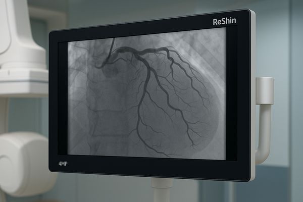
High resolution (at least 4MP–5MP) and DICOM-calibrated grayscale accuracy are critical in cardiac procedures. They ensure precise rendering of contrast flow, subtle vessel wall irregularities, and the clear depiction of fine guidewires and stents.
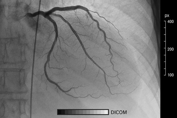
In cardiac imaging, especially during angiography and fluoroscopy, the ability to discern fine details is paramount. Resolution, often measured in megapixels (MP), directly impacts this. A higher resolution means more pixels are used to construct the image, resulting in sharper and more detailed visuals. For cardiac procedures, displays offering at least 4-megapixel to 5-megapixel resolution2 are generally recommended. This level of detail allows cardiologists to accurately visualize the subtle nuances of coronary arteries, identify the precise location and severity of stenoses (narrowings), and clearly see fine instruments like guidewires, catheters, and stents. Grayscale accuracy is equally critical. Cardiac images are predominantly monochrome, and the subtle variations in gray shades convey vital information about contrast flow dynamics, tissue density, and the structure of vessel walls. Medical displays intended for such applications must be calibrated to the DICOM (Digital Imaging and Communications in Medicine) Part 14 Grayscale Standard Display Function3. This calibration ensures that grayscale tones are rendered consistently and perceptually linearly, meaning that differences in pixel values correspond to perceivable differences in brightness. Without precise DICOM calibration, images might appear too dark or too bright, potentially obscuring subtle pathologies or leading to misinterpretation of contrast filling. This consistency is crucial not only for live imaging but also when comparing current images to prior studies. The combination of high resolution and accurate grayscale performance empowers clinicians to make more confident diagnostic and interventional decisions.
How does monitor size and layout impact workflow in cath labs?
The cath lab team works in a coordinated manner around the patient. Visibility for all team members is essential. It is important to consider how physical display characteristics affect the team’s efficiency.
Large monitors (e.g., 32-inch to 43-inch or even larger single panels) improve workflow by allowing easier long-distance viewing for the entire team. Flexible mounting on booms or mobile arms enables optimal OR integration and facilitates collaboration during procedures.
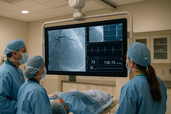
The physical size and arrangement of monitors in a cardiac catheterization lab4 significantly influence the workflow and team dynamics. Given that multiple team members—including the interventional cardiologist, nurses, and technologists—need to view the images, often from varying distances, larger screen sizes are generally preferred. Monitors in the range of 32 inches to 43 inches, or even larger single-panel displays (e.g., 55-inch or 58-inch), are common. These larger screens provide comfortable viewing from several feet away, reducing eye strain and allowing everyone in the room to follow the procedure clearly. This shared visibility is crucial for effective communication and coordination. The mounting solution for these displays is also a key consideration. In most modern cath labs, monitors are mounted on articulated ceiling booms5 or mobile C-arm systems. This allows for flexible positioning, ensuring that the screens can be optimally placed for the cardiologist performing the procedure while also remaining visible to other staff. Boom mounting keeps the floor space clear and allows the displays to be moved out of the way when not in use or during patient transfer. The ability to arrange multiple information sources on a single large display, or across a well-configured set of displays, also enhances workflow. For instance, displaying live fluoroscopy alongside reference angiograms, ECG, and hemodynamic data on one cohesive visual field prevents the need for the cardiologist to constantly shift their gaze between different, disparate screens. This integrated view6 supports quicker decision-making and a more streamlined procedural flow.
What signal and hygiene considerations apply to cath lab displays?
Cath labs are complex environments with specialized equipment. The presence of powerful X-ray systems and other electronics can create interference. It is important to address how displays cope with these challenges while maintaining sterility.
Cath labs have high EMI levels, requiring displays with well-shielded inputs and robust, low-noise internal electronics. Anti-splash, easily cleanable tempered glass surfaces are also vital for maintaining hygiene and durability in this demanding environment.
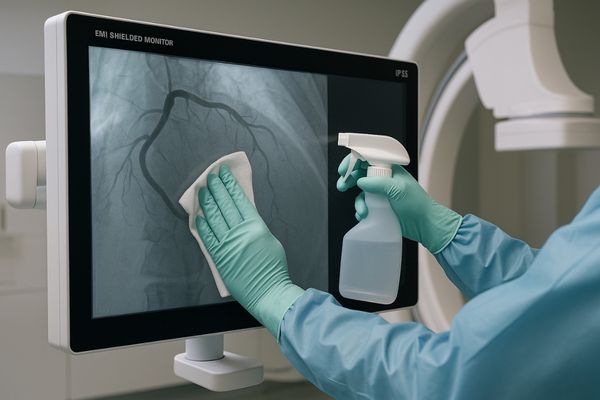
Cardiac catheterization labs present unique environmental challenges for electronic equipment, including medical displays. One major concern is Electromagnetic Interference (EMI)7. The presence of X-ray generators, C-arms, and other powerful electrical devices can create a high-EMI environment. This interference can corrupt video signals, leading to artifacts, image degradation, or even signal loss if displays are not adequately protected. Therefore, monitors designed for cath lab use must feature robustly shielded inputs and enclosures. High-quality, shielded cables are also essential. Internally, low-noise electronics and careful circuit design help to prevent the display itself from being susceptible to or generating interference. Hygiene is another paramount consideration. Cath lab procedures are invasive, and maintaining a sterile or at least very clean environment is critical to prevent infections. Displays must be designed for easy and effective cleaning and disinfection. This typically means they should have smooth, crevice-free front surfaces, often made of anti-splash tempered glass8. Such surfaces can withstand repeated wiping with harsh medical-grade disinfectants without degrading. The display housing should also be sealed to prevent liquid ingress, often achieving an IP rating (Ingress Protection)9 like IP65 on the front surface. This ensures that cleaning fluids or accidental splashes do not damage the internal electronics. Durability is also linked to hygiene; a display that can withstand rigorous cleaning protocols will have a longer operational life in the demanding cath lab setting.
Which Reshin monitors are optimized for cardiovascular imaging?
Selecting the right monitor from many options can be daunting. Specific models are often designed with particular clinical needs in mind. It is helpful to know which displays offer features tailored for heart-related procedures.
Reshin’s surgical monitors, such as the MS430PC (43-inch) and MS321PB (32-inch), are optimized for cardiovascular imaging. They feature 4MP resolution, anti-reflective (AR) glass, DICOM Part 14 support, and versatile multiple input formats, making them ideal for angiography and hybrid OR integration.
We have developed specific monitors designed to meet the rigorous demands of cardiovascular imaging environments like the cath lab and hybrid operating rooms. Our Reshin MS430PC, a 43-inch display, and the MS321PB, a 32-inch model, are prime examples. Both of these monitors offer 4-megapixel (4MP) resolution, providing the sharpness and detail necessary for visualizing fine coronary anatomy and interventional devices. This resolution strikes a good balance between detail and the ability to view images comfortably from typical distances in a cath lab. To combat glare from bright OR lights, these monitors are equipped with anti-reflective (AR) coated protective glass10. This enhances image contrast and reduces eye strain for the clinical team. Crucially, they support DICOM Part 14 grayscale calibration11. This ensures that the subtle grayscale information vital for interpreting angiograms and fluoroscopic images is displayed accurately and consistently. The ability to handle multiple input formats is another key feature. These monitors are designed to seamlessly connect to various imaging sources found in a cath lab, including C-arms, ultrasound systems, and physiological monitoring equipment. This versatility facilitates easy integration into existing setups and supports complex procedures that may require displaying information from several devices simultaneously. Their robust construction and design for easy cleaning also align with the hygiene requirements of such critical care areas.
Conclusion
Monitors for cardiac cath labs must provide high-resolution, DICOM-calibrated images with low latency. Large screens, flexible mounting, robust shielding, and hygienic design are also essential for optimal performance and safety. To upgrade your cath lab with specialized cardiac imaging displays, contact Reshin at martin@reshinmonitors.com.
-
Learning about IVUS can enhance your knowledge of advanced imaging techniques used in cardiac procedures. ↩
-
Understanding the significance of high-resolution imaging can enhance diagnostic accuracy in cardiac procedures. Explore this resource for in-depth insights. ↩
-
Learn about DICOM calibration to ensure accurate grayscale rendering, crucial for interpreting cardiac images effectively. ↩
-
Explore this resource to understand how to optimize the design of a cardiac catheterization lab for better workflow and team dynamics. ↩
-
Learn how articulated ceiling booms enhance monitor placement and flexibility in medical environments, improving team collaboration. ↩
-
Discover the advantages of an integrated view in medical imaging, which can lead to faster decision-making and improved patient outcomes. ↩
-
Understanding EMI’s impact on medical equipment is crucial for ensuring reliable performance in sensitive environments like cath labs. ↩
-
Discover how anti-splash tempered glass enhances hygiene and durability in medical settings, vital for maintaining a sterile environment. ↩
-
Learn how IP ratings ensure the durability and safety of medical displays in challenging environments, crucial for infection control. ↩
-
Discover how AR coated glass enhances image quality and reduces eye strain in medical environments. ↩
-
Learn about DICOM Part 14 calibration to appreciate its role in ensuring accurate medical image interpretation. ↩


