In the operating room, missing a subtle detail can lead to serious complications. Standard displays often lack the contrast needed to reveal critical tissue variations, creating unnecessary risk.
High-contrast monitors are essential for modern surgery because they enhance the recognition of fine details by revealing subtle tissue differences, improving the visualization of critical structures, and helping to minimize errors. This leads to more precise surgical decisions and better patient outcomes.
The ability to see clearly is fundamental to surgical success. High-contrast displays1 are not just an upgrade; they are a necessary tool for achieving the level of precision required in today’s complex medical procedures. This article will examine how superior contrast performance2 directly translates into enhanced detail recognition, safer surgeries, and a new standard of care in the operating room.
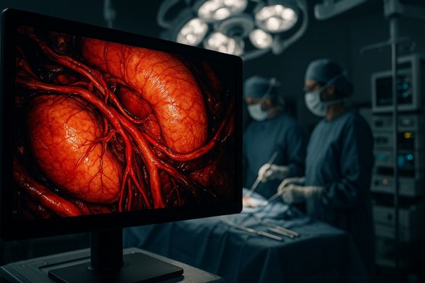
High contrast displays reveal subtle tissue differences crucial for surgical decisions
A surgeon’s decision often depends on their ability to see the faintest visual cues. A low-contrast monitor can obscure these details, leading to uncertainty at critical moments.
High-contrast displays make subtle differences in tissue color and texture clearly visible. This enhanced differentiation allows surgeons to accurately identify anatomical boundaries and make more confident, precise decisions during procedures.

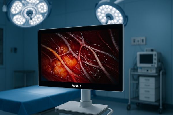
In any surgical procedure, distinguishing between different types of tissue is a fundamental task. I have consistently found that high-contrast displays3 are indispensable for this purpose. They help surgeons detect slight variations in color and texture that are essential for accurately identifying tissue boundaries. For example, the line between a tumor and healthy surrounding tissue can be incredibly subtle. A high-contrast monitor, with a contrast ratio often exceeding 1400:1, makes this boundary much easier to discern. This allows for more complete removal of diseased tissue while preserving healthy structures. Our MS275P – 27" 4K Surgical Monitor is engineered to provide this level of clarity. It ensures that the visual differences between muscle, fat, and connective tissue are not just visible, but obvious. This capability reduces surgical ambiguity and directly supports better clinical judgments, which is paramount for patient safety.
| Visual Cue | Low-Contrast Display | High-Contrast Display |
|---|---|---|
| Tissue Boundaries | Blurred, difficult to distinguish | Sharp, clearly defined |
| Color Gradations | Washed out, appear as a single color | Rich, shows subtle changes in shade |
| Surface Texture | Flat, lacks depth information | Detailed, reveals fine surface irregularities |
| Small Blood Vessels | May blend into surrounding tissue | Stand out clearly against the background |
Enhanced contrast improves visualization of blood vessels, lesions, and critical structures
During delicate surgery, small but vital structures like nerves and blood vessels can be nearly invisible on a standard screen. Damaging them can have devastating consequences for the patient.
Enhanced contrast makes fine anatomical structures such as blood vessels, nerves, and small lesions stand out with exceptional clarity. This improved visibility is essential for precise dissection and helps prevent inadvertent injury during surgery.
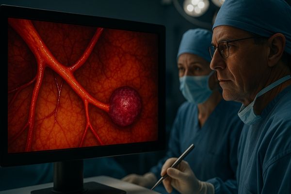
The ability to clearly see and navigate around critical structures is a cornerstone of safe surgery. High-contrast technology plays a direct role here. By increasing the visual difference between the lightest whites and the darkest blacks, these monitors make fine lines and structures pop out from the surrounding tissue. This is especially important in minimally invasive surgery, where the surgeon’s entire perception of the surgical field is mediated by a camera and a screen. A clear view of a small artery or nerve can mean the difference between a successful procedure and a serious complication. I have seen how the MS321PB – 32" 4K Surgical Monitor improves the visualization of these structures. The high contrast ensures that even the smallest vessels are not lost in the visual noise. It also allows for the accurate assessment of lesions, helping surgeons determine their size, shape, and relationship to nearby anatomy with greater confidence. This level of detail recognition reduces risk and supports better patient outcomes.
High contrast technology minimizes errors in distinguishing healthy from diseased tissue
A primary challenge in surgical oncology is ensuring every cancerous cell is removed. A standard monitor can make malignant and healthy tissue look dangerously alike, increasing the risk of incomplete resection.
High-contrast technology significantly lowers the risk of error when distinguishing between healthy and diseased tissue. By accentuating subtle visual markers, these monitors help surgeons achieve more complete tumor removal while maximally preserving healthy tissue.
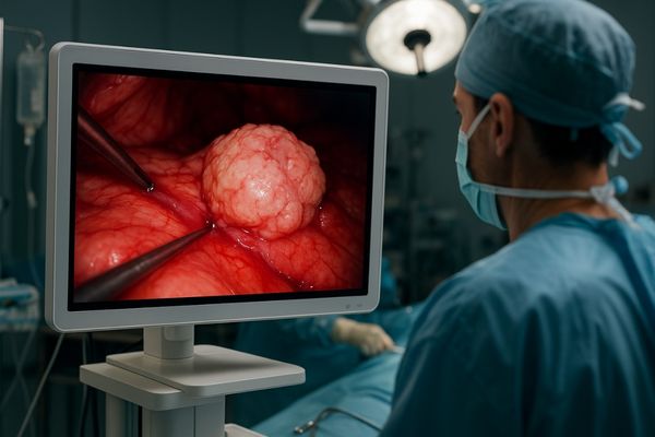
In procedures like cancer surgery, the precise performance of the monitor is directly linked to the patient’s long-term prognosis. The goal is to remove the entire tumor along with a small margin of healthy tissue, but no more than necessary. This task depends almost entirely on visual differentiation. High-contrast displays are critical in this context. I believe monitors like our MS247SA – 24" FHD Endoscopic Monitor are vital tools for surgeons because they combine high contrast with excellent color fidelity. This ensures that the subtle discoloration or textural changes that mark a tumor’s edge4 are clearly visible, even under challenging endoscopic lighting. This improved detail recognition reduces the chance of leaving malignant cells behind, which can lead to recurrence. It also helps prevent the unnecessary removal of healthy, functional tissue. By minimizing these types of errors, high-contrast technology directly contributes to more effective treatment and improved quality of life for the patient.
| Surgical Task | Impact of Low Contrast | Impact of High Contrast |
|---|---|---|
| Margin Assessment | Ambiguous tumor edges, risk of incomplete removal | Clear demarcation of margins, higher resection success |
| Lymph Node Identification | Nodes blend with fatty tissue, can be missed | Nodes are more distinct, aiding in accurate staging |
| Tissue Plane Dissection | Difficult to follow, increased risk of tissue damage | Planes are well-defined, enabling precise dissection |
| Instrument Guidance | Poor depth perception, risk of error | Improved depth cues, safer instrument handling |
Consistent contrast performance ensures reliable imaging during long procedures
A monitor’s image quality can degrade over the course of a long surgery as components heat up. This fluctuation in contrast creates visual inconsistency when surgeons need absolute reliability.
Consistent contrast performance guarantees that image quality remains stable and reliable throughout prolonged surgical procedures. Monitors built to maintain their contrast levels ensure that visual information is dependable from start to finish, preventing misinterpretation.
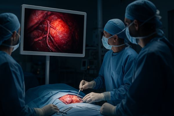
For a surgeon standing for hours, visual fatigue5 is a real problem, and an inconsistent display only makes it worse. If the contrast and brightness levels drift over time, the surgeon’s eyes are forced to constantly readjust. This is not only fatiguing but can also lead to a loss of confidence in the visual information being presented. True professional-grade surgical monitors are designed to solve this issue. They use high-quality components and advanced thermal management to ensure stable performance. Furthermore, they are often calibrated to a medical standard like DICOM Part 146, which ensures that grayscale levels are perceived consistently. This principle of consistency is critical. Our MS550P – 55" 4K Surgical Monitor, for instance, is built for endurance. It delivers uniform luminance and stable contrast from the first incision to the last suture. This reliability means surgeons can trust that the image they see in the third hour is just as accurate as the one they saw in the first, allowing them to remain focused and confident.
Reshin’s high-contrast solutions define new standards for surgical imaging clarity
Surgeons require more than just a clear picture; they need a visual instrument that provides a decisive clinical advantage. Many off-the-shelf displays fail to meet this high standard.
Our high-contrast solutions are setting new benchmarks for clarity in surgical imaging. We achieve this by integrating high contrast ratios with superior color accuracy, 4K resolution, and intelligent design to enhance surgical precision and confidence.
We believe that setting a new standard for clarity involves a holistic approach. It is not enough to excel in a single specification. Our philosophy is to combine multiple cutting-edge technologies to create a truly superior visual experience. High contrast7 is a core pillar of this philosophy, but it works in concert with other features. For example, a wide color gamut8 ensures that the enhanced contrast is applied to a true-to-life image, making tissue differentiation even more precise. High resolution provides the pixel density to render the fine details that high contrast reveals. As we look to the future, we are actively innovating in this area. We envision that the next generation of high-contrast solutions, such as our MD120C – 12MP High-Precision Diagnostic Monitor, may integrate AI to automatically enhance the visual differences between tissue types in real-time. This commitment to integrated, intelligent design is how we continue to push the boundaries of what is possible in surgical imaging.
Conclusion
High-contrast displays are fundamental to modern surgical imaging. They reveal critical details, minimize errors by improving tissue differentiation, and provide the reliable clarity needed for precision and improved patient outcomes. To enhance your surgical procedures with high-contrast medical displays, contact Reshin at martin@reshinmonitors.com.
-
Explore how high-contrast displays enhance surgical precision and improve patient outcomes. ↩
-
Learn about the critical role of superior contrast in ensuring safer and more effective surgeries. ↩
-
Explore how high-contrast displays enhance surgical precision and improve patient outcomes. ↩
-
Learn about the importance of accurately identifying a tumor’s edge to improve surgical success and patient recovery. ↩
-
Understanding visual fatigue can help improve surgical performance and monitor design. ↩
-
Exploring DICOM Part 14 will enhance your knowledge of medical imaging standards and their impact on surgical accuracy. ↩
-
Exploring this link will deepen your understanding of how high contrast enhances visual clarity and detail. ↩
-
This resource will explain the significance of a wide color gamut in achieving true-to-life images and enhancing visual experiences. ↩


