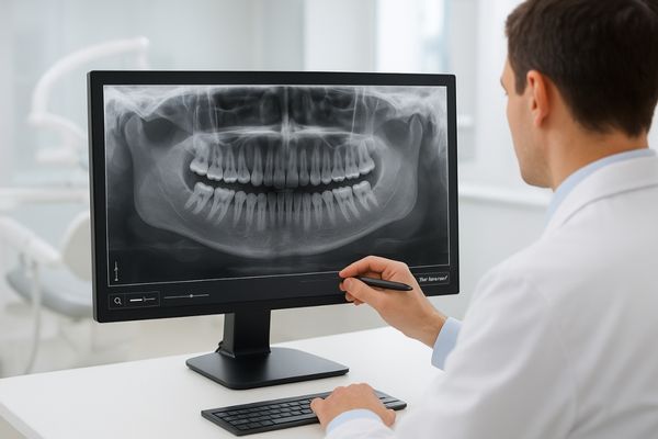
Making a diagnosis from a dental X-ray on a consumer monitor is risky. Subtle signs of caries or bone loss can be lost in poor contrast, leading to missed diagnoses.
Dental imaging systems rely on precise visualization of scans and impressions. Medical-grade displays provide the calibrated resolution, grayscale accuracy, and high brightness needed to reveal subtle details, empowering confident diagnostic decisions and enhancing patient care in modern digital dentistry.
Digital dentistry has transformed how oral health is diagnosed and treated. From intraoral X-rays and cone-beam computed tomography (CBCT) to 3D intraoral scanners, clinicians now rely on a suite of powerful imaging tools. The critical link between this advanced technology and the dentist’s diagnostic insight is the display monitor. Unlike consumer screens, medical-grade displays1 are specifically engineered to render dental images with absolute fidelity. They ensure that every subtle variation in grayscale and every nuance of color is accurately presented, which is essential for tasks ranging from identifying incipient caries to planning complex implant surgeries. By meeting rigorous standards for calibration, brightness, and consistency, these displays provide the reliability needed in a clinical setting.
Importance of Medical-Grade Displays in Dental Imaging
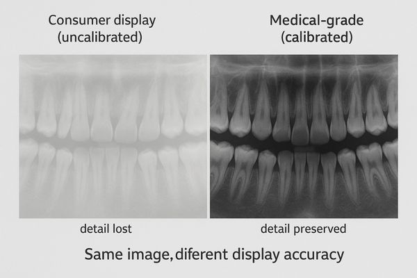
Using a standard office monitor for dental diagnostics is like using a magnifying glass with a smudged lens. Vital clinical details can be obscured, compromising patient care.
Medical-grade displays are crucial for rendering high-fidelity dental images, from X-rays to 3D models. Their precise calibration and high luminance support accurate pathology detection by maintaining uniform contrast and detail visibility in clinical environments.
The role of a display in a modern dental practice is no longer auxiliary; it is central to the diagnostic process. Medical-grade displays are purpose-built to visualize the full range of dental imaging modalities with uncompromising accuracy. Whether interpreting intraoral radiographs, panoramic scans, CBCT volumes, or digital impressions from an intraoral scanner, the monitor must faithfully reproduce the source data. These displays are engineered with factory-calibrated DICOM Grayscale Standard Display Function2 (GSDF) curves to ensure perceptual consistency in grayscale images. Furthermore, they provide high luminance and contrast to counteract the bright ambient lighting typical of a dental operatory. This ensures that subtle details remain visible and are not washed out. A quality 1MP display like the MD10C serves as a reliable entry point for viewing patient information and secondary images, establishing a baseline of quality that is essential for building a fully digital workflow.
Grayscale and Color Accuracy for Dental Diagnostics
Relying on a display with poor color or grayscale can lead to critical errors. A faint lesion might blend into the background, or an incorrect shade match can ruin a restoration.
Medical monitors offer superior 10- to 12-bit depth for thousands of gray levels and wide color gamuts for digital impressions. This expanded range reveals subtle lesions in radiographs and ensures accurate color in CAD/CAM workflows.
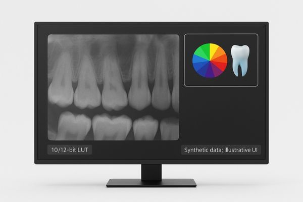
The diagnostic power of a dental image is only as good as the display it is viewed on. Medical-grade monitors vastly outperform their consumer counterparts by supporting a much wider range of grayscale and color. For grayscale-dependent modalities like bitewing and panoramic X-rays, they typically offer a 10- or 12-bit lookup table (LUT), capable of displaying up to 4,096 distinct shades of gray. This expanded depth, combined with high contrast ratios of 1000:1 or more, is essential for visualizing subtle features like early-stage enamel demineralization or fine changes in bone trabeculation. For color-critical applications3 such as digital impressions and CAD/CAM shade matching, displays like the MD22CA provide accurate color reproduction, often covering over 72% of the NTSC color gamut. This precision ensures that the virtual model on the screen is a true representation of the patient’s dentition, which is fundamental for creating well-fitting and aesthetically pleasing restorations.
DICOM Calibration and Consistent Image Quality
An uncalibrated monitor is unpredictable. The same X-ray can look different from one day to the next, eroding diagnostic confidence and consistency across a practice.
Medical displays adhere to the DICOM Part 14 standard, ensuring consistent luminance response aligned with human vision. Built-in sensors and automatic calibration maintain this standard, significantly improving lesion detection and diagnostic reliability.
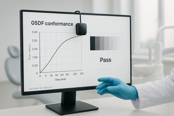
Consistency is a cornerstone of quality care in dentistry. To achieve this in digital imaging, diagnostic displays must conform to the DICOM Part 14 Grayscale Standard Display Function (GSDF)4. This standard ensures that the monitor’s luminance response is perceptually linear, meaning that changes between grayscale levels are equally noticeable to the human eye. This is not a "set it and forget it" feature. Over time, all displays degrade. Medical-grade monitors address this with built-in calibration sensors and sophisticated LUTs that automatically adjust the output to maintain GSDF compliance. Some models feature dynamic LUT technology5 that continually re-calibrates luminance in real time. This automated quality control, found in models like the high-brightness MD26GA, is proven to improve the detection of caries and other pathologies, particularly in the challenging, brightly lit conditions of a dental clinic. It ensures that every clinician in a practice sees the same image in the same way, every time.
Resolution Needs Across Dental Imaging Modalities
Using a low-resolution screen for a high-detail CBCT scan is a critical mismatch. It forces excessive zooming and panning, making it easy to lose context and miss important findings.
Dental modalities require varying resolutions. While 1-2MP is sufficient for intraoral films, panoramic and CBCT scans benefit from 3-5MP displays to show full-arch detail and multiple views without compromising clarity.
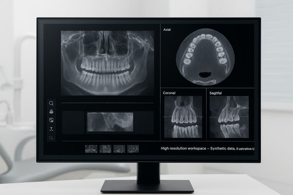
Different dental imaging modalities generate data with varying levels of detail, imposing different demands on display resolution. For instance, intraoral radiographs6 like bitewings are relatively small images, but they require a high pixel density (a small pixel pitch) to resolve the fine details of interproximal caries. A 2MP monitor is often a good match for this task. However, larger datasets from panoramic X-rays7 or CBCT scans benefit significantly from higher resolutions. A 3MP display, such as the MD32C, allows a clinician to view an entire panoramic arch or multiple CBCT cross-sections side-by-side with exceptional clarity, minimizing the need to constantly zoom and pan. For advanced multi-modality workflows, even larger 4K monitors can be used to display X-rays, 3D models, and patient photos concurrently. The key is to match the display’s resolution and size to the primary imaging tasks to optimize diagnostic efficiency and accuracy.
| Imaging Modality | Common Resolution Need | Key Requirement |
|---|---|---|
| Intraoral X-ray (Bitewing) | 1–2 MP | High pixel density (small pitch) |
| Panoramic Radiograph | 2–4 MP | Wide format, full-arch detail |
| CBCT (Cone-Beam CT) | 3–5 MP+ | Ability to display multiple planes |
| Intraoral 3D Scan / Photos | 2–4 MP | High color accuracy |
Latency and Real-Time Display in Dental Applications
During a guided implant surgery, display lag can be disastrous. A delay between the surgeon’s movement and the on-screen feedback can lead to inaccurate placement.
While many dental images are static, displays used for live scanning or guided surgery require low latency. Medical monitors offer fast response times and high refresh rates to ensure on-screen feedback is instantaneous, preventing motion blur and delay.
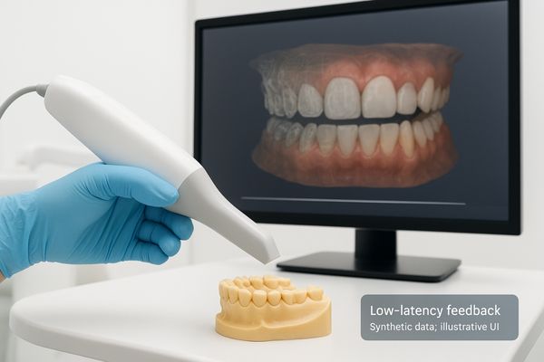
Although much of dental image review involves static images, some advanced applications depend on real-time video feedback8 where display performance is critical. During procedures like guided implant placement or live intraoral scanning, any perceptible delay—or latency—between the action and the on-screen representation can disrupt the workflow and compromise precision. Medical displays are engineered for low latency9, with typical response times in the range of 10-30 milliseconds and refresh rates of 60 Hz or higher. This ensures that motion appears smooth and free of distracting blur or judder. In surgical contexts, an input lag of even less than 100 milliseconds can be critical for maintaining hand-eye coordination. By using fast processors and modern high-speed interfaces like DisplayPort, a calibrated color diagnostic display like the MD45C ensures that what the clinician sees on the screen is a true real-time reflection of the instrument’s position or the scanner’s progress.
Brightness and Ambient Light Adaptation in Clinics
Viewing an X-ray in a bright dental office can wash out the image. The glare from overhead lights can easily hide the subtle gray shades that indicate early decay.
Dental clinics are bright environments, requiring displays with very high peak luminance (often 1000 cd/m² or more) to preserve contrast. Anti-glare coatings and ambient light sensors further ensure that images remain clear and diagnostically useful.
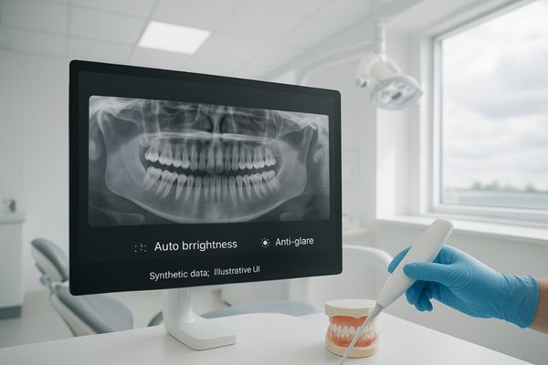
Dental operatories are typically much brighter than the dark reading rooms used in radiology. This high level of ambient light poses a significant challenge, as it can reflect off the screen and drastically reduce perceived image contrast. To combat this, medical-grade displays provide very high peak luminance10, often reaching 1000 to 2000 cd/m². This allows them to effectively "punch through" the ambient light, ensuring that grayscale details and subtle features remain visible. Studies have confirmed that the ability to detect carious lesions drops sharply in brightly lit environments. While best practice is to dim task lighting for diagnostic review, high-brightness monitors act as a critical safeguard. Many models, such as theMD46C, also incorporate highly effective anti-glare coatings11 and automatic ambient light sensors that adjust brightness to match the room’s conditions, guaranteeing a clear and consistent view.
System Compatibility and Workflow Integration
A new display that cannot connect to the existing imaging workstation is useless. Incompatibility can halt the entire digital workflow, causing delays and frustration.
Medical-grade displays are designed for seamless integration. They support standard interfaces, are validated with major PACS software, and include on-board QC tools for simplified management and regulatory compliance within a connected digital practice.
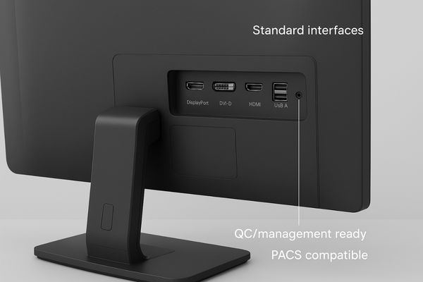
For a display to be a valuable asset in a dental practice, it must integrate seamlessly into the existing digital imaging workflow. This means more than just having the right video port. Medical-grade displays are designed for deep compatibility. They support standard interfaces like DisplayPort, DVI, and HDMI to connect easily with any modern imaging workstation. Their firmware and drivers are extensively tested with major PACS and DICOM viewer software to ensure that images and calibration data are transferred correctly without corruption. Furthermore, they often include on-board quality control (QC) software12 suites, allowing practice administrators to remotely manage calibration schedules and log compliance data for regulatory purposes. Some models even offer advanced features like picture-in-picture or split-screen modes, enabling the simultaneous comparison of different image types. This commitment to compatibility ensures the display is a reliable, manageable, and compliant endpoint in a sophisticated clinical ecosystem.
Conclusion
By providing calibrated accuracy, high brightness, and reliable consistency, medical-grade displays are an essential component of modern digital dentistry, empowering professionals to diagnose with confidence and elevate the standard of care.
👉 For personalized advice on dental imaging displays that meet the highest standards, contact Martin at martin@reshinmonitors.com — we’ll guide you to the best solution.
-
Learn about the significance of medical-grade displays in ensuring accurate dental imaging and enhancing diagnostic precision. ↩
-
Learn about DICOM GSDF and its critical role in ensuring consistent and accurate grayscale image reproduction in dental diagnostics. ↩
-
Learn about the importance of color accuracy in dental imaging and how it affects treatment quality and patient satisfaction. ↩
-
Understanding GSDF is crucial for ensuring consistent and high-quality digital imaging in dentistry. ↩
-
Explore how dynamic LUT technology enhances image quality and consistency in medical displays, vital for accurate diagnostics. ↩
-
Understanding intraoral radiographs is crucial for dental professionals to enhance diagnostic accuracy and patient care. ↩
-
Exploring panoramic X-rays can provide insights into their advantages for comprehensive dental assessments and treatment planning. ↩
-
Understanding real-time video feedback is crucial for improving surgical precision and workflow efficiency. ↩
-
Exploring the significance of low latency can enhance your knowledge of display technology in critical medical applications. ↩
-
Understanding high peak luminance is crucial for ensuring optimal image quality in bright environments, enhancing diagnostic accuracy. ↩
-
Exploring anti-glare coatings can reveal how they enhance visibility and comfort, making them essential for effective diagnostics. ↩
-
Learn about the importance of QC software in maintaining image integrity and regulatory compliance. ↩


