A hospital can invest millions in a PACS, but the system is useless without the right screen. A poor display can hide critical details, making the entire digital chain ineffective.
High-resolution displays are the foundation of accurate diagnostic imaging within PACS. This article explains how DICOM compliance, multi-modality integration, ergonomic design, and system compatibility work together to create a reliable and efficient diagnostic environment.
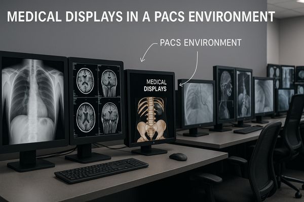
The Picture Archiving and Communication System (PACS) is the digital heart of modern radiology. It has revolutionized how we store, retrieve, and view medical images. However, the final, and perhaps most critical, component in this chain is the medical display1. This screen is the window through which clinicians interpret complex visual data. The quality of this window directly impacts diagnostic accuracy2, workflow efficiency, and even the well-being of the medical professional. Let’s explore the key roles these displays play.
High-Resolution Displays Support Accurate Image Interpretation
Low-resolution screens make it easy to miss tiny, critical details in a medical scan. This can lead to diagnostic delays or errors. High-resolution displays ensure every pixel tells a story.
High-resolution displays clearly present the fine details in CT, MRI, and X-ray images within PACS systems. This clarity forms the essential foundation for any accurate diagnosis.
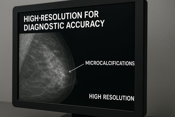
In medical imaging, resolution is about more than just a sharp picture; it is about diagnostic data3. A higher resolution means more pixels are used to display the image, allowing for the visualization of smaller anatomical structures. For a radiologist examining a mammogram, this could mean the difference between spotting a tiny cluster of microcalcifications and missing an early sign of cancer. For a neurologist reviewing an MRI, it means seeing the subtle tissue changes that indicate a stroke. Lower-resolution displays force the user to constantly zoom and pan, which disrupts the reading flow and increases the risk of overlooking something outside the magnified area. High-resolution displays, such as the MD120C – 12MP High-Precision Diagnostic Monitor with AI Calibration, provide a complete and detailed view of the entire image at once. This comprehensive perspective is fundamental to building diagnostic confidence and ensuring that clinical decisions are based on the most complete visual information available.
| Resolution | Common Modalities | Key Benefit |
|---|---|---|
| 2MP / 3MP | CT, MRI, General Radiography | Excellent for clinical review and general diagnostic tasks. |
| 5MP | Mammography, Tomosynthesis | Required standard for resolving microcalcifications. |
| 8MP / 12MP | Multi-modality Fusion, Chest X-ray | Allows viewing of multiple large images or a single giant image without compromise. |
DICOM Compliance Ensures Consistency Across Viewing Stations
An image looks bright and clear in the radiology department but dark and muddy on a clinician’s review station. This inconsistency creates confusion and undermines trust in the diagnosis.
DICOM compliance guarantees that luminance and grayscale are consistent across all viewing stations and departments. This standardization is critical for reducing the risk of diagnostic errors caused by display variability.
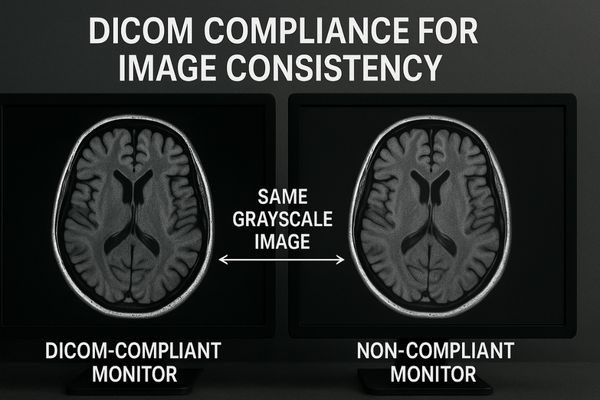
The DICOM (Digital Imaging and Communications in Medicine) standard is the universal language of medical imaging. DICOM Part 14 specifically addresses the grayscale display function. It defines a precise curve for how pixel values in an image should be rendered as light on a screen. Why is this so important? It ensures that a specific grayscale value always corresponds to the same level of brightness, regardless of the monitor being used. This principle, known as perceptual consistency4, is vital. It means a radiologist, a surgeon, and a referring physician can all look at the same image on different DICOM-compliant displays and see the exact same clinical information. Consumer-grade monitors cannot achieve this; their brightness fluctuates with age and temperature. Medical displays like the MD33G – 3MP Grayscale Diagnostic Monitor use built-in sensors and automated calibration software to constantly check and adjust their output, ensuring they remain perfectly aligned with the DICOM standard5 throughout their lifespan. This removes display variation as a source of error.
Multi-Modality Imaging Integration Enhances Diagnostic Workflow
A clinician needs to compare a patient’s new CT scan with a previous PET scan. They waste valuable time opening different applications and dragging windows across multiple screens. This is inefficient.
Multi-modality integration allows clinicians to simultaneously compare results from different exams on a single, unified display. This significantly improves diagnostic efficiency and speeds up the decision-making process.
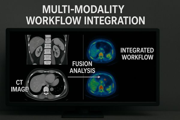
Modern medicine rarely relies on a single type of imaging. A patient’s diagnosis often involves correlating information from multiple sources, such as CT for anatomical structure, MRI for soft tissue detail, and PET for metabolic function. A traditional setup with multiple disparate monitors makes this comparison process cumbersome and inefficient. Clinicians lose time and focus switching their attention between screens. Multi-modality displays6 were created to solve this problem. These are often large, single-panel displays that can seamlessly show images from two or more different sources at the same time using functions like Picture-by-Picture (PBP) or Picture-in-Picture (PIP). For example, a single workstation equipped with a monitor like the MD46C – Dual-screen Diagnostic Monitor (Single Panel) can replace a cluttered desk with two separate monitors. This integrated view allows for faster correlation of findings, a more holistic understanding of the patient’s condition, and a dramatically streamlined diagnostic workflow. The time saved translates directly into faster report turnaround and improved patient care.
Ergonomic Features Improve Radiologist Reading Comfort
Radiologists spend their entire day staring at screens, leading to eye strain and neck pain. This physical discomfort reduces their stamina and can degrade their diagnostic performance over time.
Ergonomic features like adjustable monitor height, customizable tilt, and low blue light technology help alleviate physical fatigue during long reading sessions. This directly boosts radiologist productivity and well-being.
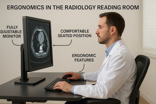
The work of a radiologist is visually and cognitively demanding. They can spend eight hours or more per day in a darkened room, interpreting hundreds of complex images. This sustained effort can take a significant physical toll. Poor ergonomics can lead to musculoskeletal issues, while constant screen viewing contributes to Computer Vision Syndrome (CVS)7, with symptoms like eye strain, dryness, and headaches. Medical displays designed with advanced ergonomic features can mitigate these risks. A fully adjustable stand that allows for height, tilt, swivel, and pivot adjustments lets each user position the screen perfectly for their posture. Technologies like flicker-free backlights and dedicated low-blue-light modes reduce the principal causes of eye fatigue. Even screen surface treatments that minimize glare and reflections play a role. A product like the MD32C – 3MP Diagnostic Monitor incorporates these features to create a more comfortable and sustainable reading environment. A comfortable radiologist is a more focused, efficient, and accurate radiologist.
| Ergonomic Feature | Problem It Solves | Benefit for Radiologist |
|---|---|---|
| Adjustable Stand | Poor posture, neck/back pain | Personalized viewing angle, reduced musculoskeletal strain. |
| Flicker-Free Backlight | Sub-perceptual screen flicker | Reduces eye strain and headaches during long sessions. |
| Low Blue Light Mode | Disruption of circadian rhythm | Improves comfort for late-night reading, can help sleep patterns. |
| Anti-Glare Surface | Distracting reflections | Maintains image clarity and reduces visual effort. |
Network and System Compatibility Streamline PACS Deployment
A hospital buys new displays, but they fail to connect properly to the existing workstations or PACS software. The IT department spends weeks troubleshooting, delaying the upgrade and causing frustration.
Good compatibility with existing hospital networks and major PACS platforms ensures a smooth and rapid system deployment. This, in turn, helps to lower long-term maintenance costs and complexity.
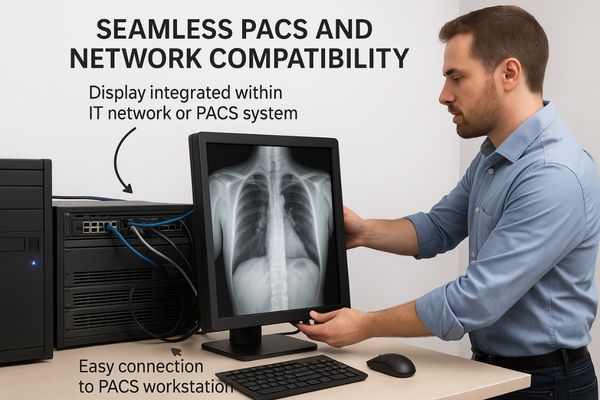
A medical display does not operate in isolation; it is a networked endpoint within a complex hospital IT infrastructure. A smooth deployment depends entirely on its ability to integrate seamlessly with existing hardware and software. This compatibility extends across several areas. First, it requires support for standard video interfaces like DisplayPort and HDMI to connect to a wide range of workstations and graphics cards. Second, the display’s drivers and control software must be compatible with the hospital’s operating systems, typically various versions of Windows. Most importantly, the display’s remote calibration and Quality Assurance (QA) software8 must be able to communicate effectively with the central PACS server. This allows the hospital IT or medical physics team to monitor, manage, and verify the performance of every display on the network from a single location. Choosing a display like the MD26GA – 2MP Diagnostic Monitor, which is designed and tested for broad compatibility with major PACS vendors, simplifies this entire process. It means less time spent on integration and more time spent on patient care.
Conclusion
A medical display is the critical endpoint of any PACS. Its resolution, consistency, usability, and compatibility directly influence diagnostic accuracy, workflow efficiency, and the clinician’s own well-being.
📧 Looking to optimize your PACS setup with high-performance medical displays? Contact Martin at martin@reshinmonitors.com for tailored solutions from Reshin.
-
Understanding the role of medical displays can enhance diagnostic accuracy and improve workflow efficiency in healthcare. ↩
-
Exploring this topic reveals how crucial accurate diagnostics are for patient care and treatment success. ↩
-
Learn about the critical role of diagnostic data in making informed clinical decisions and improving patient care. ↩
-
Understanding perceptual consistency is crucial for ensuring accurate medical imaging across different displays, enhancing diagnostic reliability. ↩
-
Exploring the DICOM standard will provide insights into its role in medical imaging, ensuring consistency and accuracy in diagnostics. ↩
-
Explore this link to understand how Multi-modality displays enhance diagnostic efficiency and improve patient care. ↩
-
Understanding CVS is crucial for radiologists to prevent eye strain and maintain productivity. ↩
-
Understanding QA software is crucial for ensuring the reliability and performance of medical displays in healthcare settings. ↩