Precision medicine relies on subtle image data. Standard screens can hide these details, compromising personalized care. High-fidelity medical displays ensure every critical clue is visible for accurate treatment.
Medical displays significantly impact precision medicine by enabling accurate interpretation of complex imaging data. They support multimodal visualization, ensure color-critical tissue differentiation, and facilitate collaboration. This high-fidelity view is essential for developing personalized, data-driven treatment strategies for each patient.
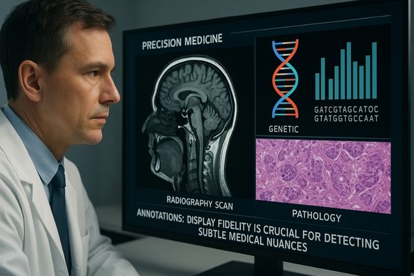
Precision medicine is not just about genetics or data algorithms; it is about how that information is visually presented to the clinician. The display is the final gateway where complex data becomes actionable insight. In this article, we will explore the specific ways high-quality medical displays1 are not just helpful, but absolutely essential to the success of precision medicine, from initial diagnosis to collaborative treatment planning. Let’s look closer at how this technology makes personalized care2 possible.
High-Fidelity Displays Enable Accurate Image Interpretation
In precision medicine, a tiny anomaly can change everything. Standard displays can blur these subtle clues, leading to missed findings. High-fidelity displays ensure every detail is seen with clarity.
High-fidelity displays enable accurate image interpretation by providing the exceptional resolution, brightness, and contrast needed to see subtle pathologies. This clarity allows clinicians to confidently distinguish between benign and malignant features, which is fundamental to creating a precise and effective treatment plan.
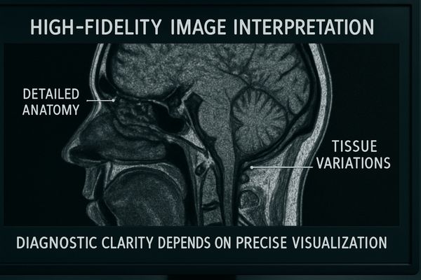
In the context of precision medicine, "high-fidelity3" goes beyond simple screen clarity. It refers to the display’s ability to render an image without losing any information from the original digital file. This involves several technical aspects. High resolution, like that found in 4K or 8K displays, provides the pixel density to visualize microscopic structures. High bit depth allows the monitor to show thousands of shades of gray, which is critical for seeing subtle density variations in tissues. Finally, strict adherence to the DICOM GSDF4 ensures that these grayscale values are perceptually linear and consistent. In precision medicine, a radiologist might be looking for a slight change in tissue texture that indicates a specific biomarker. A low-fidelity display would average out these pixels, making the texture invisible. A high-fidelity screen preserves this data, turning a simple image into a rich source of information for personalized treatment. Our MD50C – 5MP Color Mammography Monitor is designed for this purpose, providing the fidelity needed to detect the earliest, most subtle signs of disease.
Multimodal Visualization Supports Data-Driven Decisions
Clinicians juggle data from radiology, pathology, and genomics. Constantly switching between systems is slow and confusing. Multimodal displays bring all this information together for a single, comprehensive view.
Multimodal visualization supports data-driven decisions by fusing different data types—like PET/CT scans, MRI, and genomic markers—onto a single display. This integrated view allows clinicians to see the relationships between anatomical structures and metabolic activity, leading to more personalized and effective treatment plans.
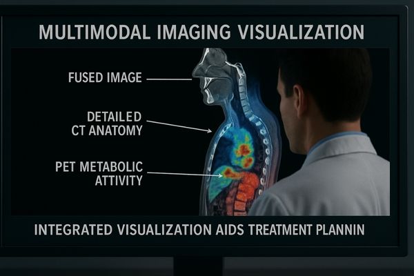
Precision medicine thrives on synthesis. It is about connecting information from different sources to build a complete patient profile. Multimodal visualization5 is the practical application of this principle. Techniques like PET/CT fusion6 are a great example. The CT scan provides a detailed anatomical map in grayscale, while the PET scan overlays metabolic activity in color. A clinician needs a display that can render both data types with perfect accuracy on the same screen. This requires a monitor with a hybrid gamma curve, capable of following the DICOM standard for the CT portion and a separate gamma curve for the color PET data. This fusion allows a doctor to see not just where a tumor is, but also how active it is, which is crucial for staging cancer and planning targeted radiation therapy. A large-format display like our MD51CHY – 34" 5MP Diagnostic Monitor provides the screen space and technical capability to view these fused datasets without compromise, turning complex data into a clear, actionable plan.
Data Integration in Precision Medicine
| Data Source | Information Provided | Role of the Display |
|---|---|---|
| Radiology (CT/MRI) | Anatomical structure, tissue density | Accurate grayscale (DICOM) rendering |
| Nuclear Medicine (PET) | Metabolic activity, cellular function | Precise color visualization of hotspots |
| Genomics/Pathology | Biomarker status, genetic mutations | Clear text and image overlay on scans |
Color Accuracy is Crucial for Tissue Differentiation
Pathologists rely on subtle color stains to identify disease. An inaccurate display can distort these colors, leading to a potential misdiagnosis. Color-calibrated medical displays render tissue stains with perfect fidelity.
Color accuracy is crucial because pathologists and surgeons use stains to differentiate tissue types. Precise color reproduction ensures that what they see on screen exactly matches what is under the microscope or in the surgical field, which is vital for accurate diagnosis and for preserving healthy tissue.
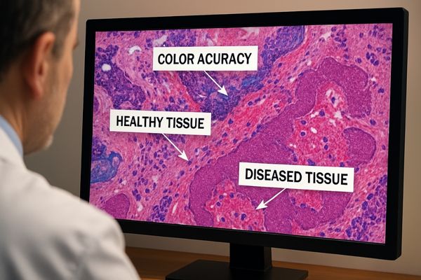
While radiology often focuses on grayscale, many pillars of precision medicine are built on color. In digital pathology, hematoxylin and eosin (H&E) stains7 color cell nuclei blue and the cytoplasm pink. The exact shade, intensity, and distribution of these colors help pathologists identify cancerous cells. If a monitor has a slight blue tint, for example, it could artificially enhance the appearance of nuclei, potentially leading to an over-grading of a tumor. The same is true in the operating room. In fluorescence-guided surgery8, a surgeon injects a dye that makes cancer cells glow a specific color (like green or red) under a special light. The surgical display must reproduce this fluorescent color with absolute precision against the background of normal tissue. Any color inaccuracy could cause the surgeon to remove too much healthy tissue or leave cancerous cells behind. The MS321PB – 32" 4K Surgical Monitor is engineered with a wide color gamut and advanced calibration tools to ensure that these critical color distinctions are always preserved.
Display Consistency Enhances Cross-Disciplinary Collaboration
A patient’s care team is spread across departments. If their displays show images differently, collaboration breaks down. Standardized, calibrated displays ensure everyone sees the exact same image, everywhere.
Display consistency enhances collaboration by providing a shared, reliable visual baseline for all members of a care team. When radiologists, surgeons, and oncologists all view images on calibrated, DICOM-compliant displays, it eliminates ambiguity and ensures that treatment decisions are based on the same accurate information.
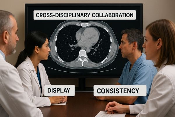
The tumor board is a cornerstone of modern cancer care and a perfect example of precision medicine in action. Here, experts from radiology, pathology, surgery, and oncology meet to create a unified treatment plan. This collaboration is only effective if everyone is looking at the same visual information. The radiologist projects a grayscale CT scan, which must be DICOM compliant9. Moments later, the pathologist displays a colorful digital slide, which must be color-accurate10. The team’s decisions depend on combining insights from both. If the main presentation display has a poor contrast ratio or inaccurate color, the discussion is compromised from the start. A standardized display environment, where every monitor from the radiologist’s desk to the conference room is calibrated to the same high standard, removes this variable. It builds a foundation of trust in the images, allowing the team to focus on their collective expertise. A large-format display like our MS550P – 55" 4K Surgical Monitor is ideal for these settings, providing the size and quality needed for the entire team to see every detail clearly.
The Role of Consistent Displays in Tumor Boards
| Specialist | Type of Image | Key Display Requirement |
|---|---|---|
| Radiologist | CT, MRI, X-ray | DICOM Part 14 Grayscale Consistency |
| Pathologist | Digital Histology Slides | sRGB/Adobe RGB Color Accuracy |
| Surgeon | Endoscopy, Laparoscopy | High Brightness, Low Latency, Color Fidelity |
AI Integration Elevates Imaging in Precision Medicine
AI algorithms can detect subtle disease markers in images. But if the clinician can’t see the AI’s findings clearly, the insight is lost. AI-ready displays make these outputs visible.
AI integration elevates imaging by using algorithms to detect patterns and quantify findings that are invisible to the human eye. A medical display then visualizes these results—such as heatmaps or boundary overlays—turning raw AI data into actionable clinical insights for more precise diagnosis and treatment.
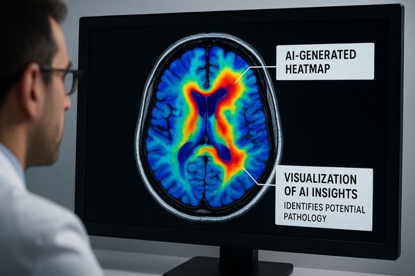
Artificial intelligence is moving from a research concept to a practical clinical tool. In precision medicine11, AI algorithms can analyze thousands of images to learn subtle patterns that predict treatment response or disease progression. For example, an AI might analyze a CT scan and produce a colored heatmap indicating the probability of malignancy for different parts of a lung nodule. The medical display’s job is to present this complex, AI-generated information in a way that helps, not hinders, the radiologist. It must render the AI’s colored overlay with precision while maintaining the integrity of the original grayscale scan underneath. This requires a display with exceptional dynamic range and fine control over brightness and color. The clinician must be able to trust that the AI’s findings are displayed clearly and without distortion, allowing them to use it as a "second reader" to confirm their own analysis. A model like our MD33G – 3MP Grayscale Diagnostic Monitor provides the stable, DICOM-compliant12 canvas needed to integrate these AI insights effectively into the diagnostic workflow.
Conclusion
Ultimately, precision medicine demands precision tools. High-quality medical displays are essential, providing the clear, consistent, and data-rich visualizations that make personalized patient care a reality.
📧 Ready to upgrade your imaging equipment for precision care? Contact Martin at martin@reshinmonitors.com to explore Reshin’s advanced medical display solutions.
-
Discover how high-quality medical displays enhance the effectiveness of precision medicine and improve patient outcomes. ↩
-
Explore the significance of personalized care in modern medicine and its impact on patient satisfaction and recovery. ↩
-
Understanding high-fidelity displays is crucial for professionals in precision medicine to ensure accurate diagnostics. ↩
-
Exploring DICOM GSDF will enhance your knowledge of image consistency and quality in medical imaging. ↩
-
Explore this link to understand how multimodal visualization enhances patient profiles and improves clinical decision-making. ↩
-
Learn about PET/CT fusion to see how it combines anatomical and metabolic data for better cancer diagnosis and treatment. ↩
-
Understanding H&E stains is crucial for grasping how pathologists identify cancerous cells through color analysis. ↩
-
Exploring fluorescence-guided surgery will reveal its significance in ensuring precise tumor removal and preserving healthy tissue. ↩
-
Understanding DICOM compliance is crucial for ensuring accurate medical imaging and effective collaboration in tumor boards. ↩
-
Exploring the significance of color accuracy in pathology can enhance your understanding of diagnostic precision and treatment planning. ↩
-
Understanding precision medicine is crucial as it highlights how AI enhances personalized treatment strategies, making healthcare more effective. ↩
-
Exploring DICOM compliance is essential for grasping how medical images are standardized, ensuring accurate AI analysis and reliable diagnostics. ↩