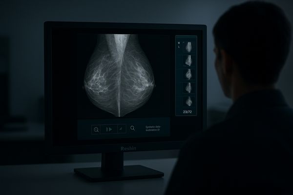
A radiologist squints at a mammogram on a standard screen. A subtle cluster of microcalcifications, a potential early sign of cancer, is lost in the screen’s poor contrast and glare.
Digital mammography demands exceptional grayscale fidelity, high luminance, and uncompromising uniformity. Reshin medical-grade monitors are engineered for this task, ensuring consistent perception, faster workflows, and reliable detection with 5MP, 8MP, and 12MP display options.
The transition from film to digital mammography, including full-field digital mammography (FFDM) and now digital breast tomosynthesis (DBT)1, has placed immense demands on display technology. The goal is to detect the earliest and most subtle signs of breast cancer, such as faint microcalcifications and low-contrast masses hidden within dense tissue. This requires a level of image fidelity that standard commercial monitors cannot provide. Medical-grade mammography displays are purpose-built to meet this challenge. They are not merely screens, but calibrated diagnostic instruments designed for absolute consistency, superior detail visualization, and optimized workflow efficiency. This article will explore the key clinical tasks in digital mammography and detail how specialized displays provide the technological foundation for confident and accurate interpretation in this critical field.
Clinical Tasks in Digital Mammography: FFDM, DBT, CAD
A radiologist toggles between multiple windows, trying to compare different DBT slices. This constant switching is slow and breaks concentration, increasing the risk of missing a subtle finding.
Mammography involves interpreting FFDM, DBT, and CAD results. Medical displays must present these varied static and dynamic images with perfect clarity to support accurate detection of masses and microcalcifications.
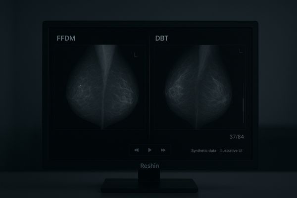
Digital mammography encompasses several distinct but related clinical tasks, each placing specific demands on the display. Full-field digital mammography (FFDM) produces high-resolution static 2D images. The primary challenge here is resolving fine microcalcifications and distinguishing low-contrast soft-tissue masses from the surrounding breast tissue. Digital breast tomosynthesis2 (DBT), or 3D mammography, acquires a series of low-dose projection images that are reconstructed into thin slices. This requires the radiologist to scroll through a "cine loop" of dozens of images, a dynamic task that demands a display capable of smooth rendering without artifacts. Finally, computer-aided detection3 (CAD) systems overlay markers onto the images to draw attention to potential areas of concern. The display must render these overlays clearly without obscuring the underlying anatomical data. A proven baseline display like the MD52G is specifically designed for these grayscale-critical tasks, with a 5MP resolution that has long been the gold standard for FFDM interpretation, ensuring no detail is lost.
Resolution & Pixel Pitch: From 5MP to 8MP/12MP
Using a lower-resolution monitor for DBT is like reading a map through a keyhole. The reader is forced to constantly pan and zoom, losing the overall anatomical context.
While 5MP is the baseline for FFDM, the massive datasets of DBT benefit from 8MP and 12MP single-head displays, which allow more images to be viewed simultaneously with fewer manipulations.
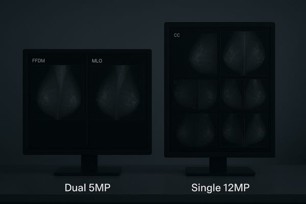
Resolution is a critical parameter in mammography. The goal is to match the display’s pixel density to the detail captured by the imaging detector. For years, a pair of 5-megapixel (5MP) monitors4 has been the established standard for FFDM. The small pixel pitch of these displays is essential for resolving individual microcalcifications. However, the advent of DBT5, which can generate hundreds of images per study, has shifted the paradigm. Radiologists must rapidly scroll through image stacks, and a larger digital canvas improves this workflow dramatically. This has driven the adoption of single-head 8MP and 12MP displays. These larger monitors allow for multiple DBT slices, prior images, or different views (like CC and MLO) to be displayed side-by-side on a single, seamless surface. This reduces the distracting "bezel gap" of dual-monitor setups and minimizes the eye and head movement required to "pan and scan" across the dataset, ultimately improving throughput and reducing reader fatigue.
Luminance, Contrast & Ambient Light Management
In a reading room with imperfect lighting, glare can wipe out subtle image details. A faint mass visible in a dark room can become invisible under normal ambient light.
High peak luminance and superior contrast ratios are essential for preserving lesion conspicuity in real-world lighting conditions. Anti-glare surfaces and brightness stabilization ensure consistent detail perception.
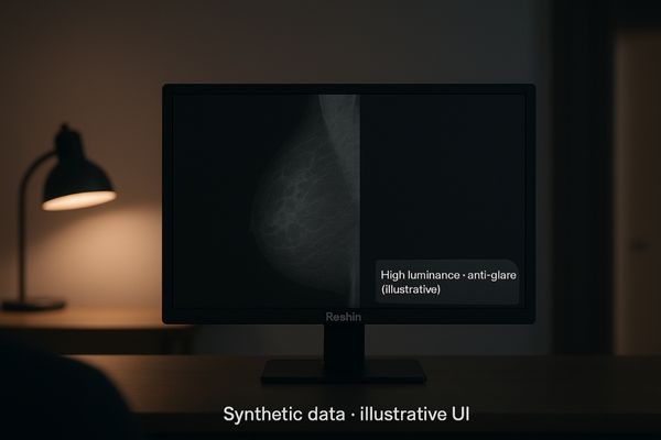
The ideal radiology reading room is perfectly dark, but in practice, ambient light from doorways, other workstations, or overhead lighting is common. This stray light can reflect off the monitor’s surface, washing out the image and reducing contrast, which is particularly dangerous in mammography where low-contrast masses are a key finding. To combat this, medical-grade mammography displays6 provide very high peak luminance, often far exceeding the minimum requirements. This high brightness allows the display to maintain excellent contrast even when ambient light cannot be fully controlled. In addition, these monitors feature advanced anti-glare and low-reflection screen surfaces that diffuse reflections without blurring the image. This ensures that the deep blacks and subtle grays that define a lesion’s margins remain conspicuous. Constant brightness control technology7 also ensures this high level of luminance is maintained consistently over the monitor’s lifespan, providing reliable performance for years.
Grayscale Fidelity & DICOM Part 14 Calibration
If a monitor is not properly calibrated, two different gray shades can appear identical. This can make it impossible to differentiate a cyst from a potentially malignant solid mass.
Hardware calibration to the DICOM Part 14 standard is non-negotiable. This ensures a perceptually linear grayscale response, which is critical for the consistent and accurate detection of subtle calcifications and masses.
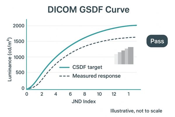
The entire practice of mammographic interpretation relies on the faithful reproduction of grayscale information. The display must present the image data in a way that is perceptually linear, meaning that equal changes in pixel values result in equally perceptible changes in brightness to the human eye. This is the purpose of the DICOM Part 14 Grayscale Standard Display Function (GSDF)8. Medical-grade mammography displays undergo rigorous hardware calibration at the factory, where an internal 12- or 14-bit look-up table (LUT) is programmed to ensure the display’s output precisely matches the DICOM curve. This is not a software adjustment; it is a fundamental characteristic of the monitor’s performance. This ensures that a radiologist can consistently differentiate between the subtle gray levels that define the edges of a mass or the texture of microcalcifications. Routine quality assurance checks, often facilitated by built-in sensors, confirm that this calibration is maintained over time, safeguarding diagnostic integrity.
Uniformity, Stabilization & Long-Term Consistency
A monitor that is brighter in the center than at the corners is a major liability. A faint lesion in a dimmer corner of the screen could be completely missed.
Panel uniformity is as critical as resolution. Advanced compensation technology ensures consistent brightness from corner to corner, while stabilization systems counteract backlight aging, guaranteeing long-term image consistency.
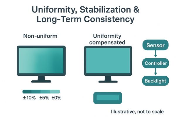
A monitor’s specifications are meaningless if they only apply to the center of the screen. In mammography, a radiologist scrutinizes every millimeter of the display. For this reason, screen uniformity is just as important as resolution or brightness. Medical-grade displays incorporate sophisticated Uniformity Equalizer technology9, which measures and corrects for brightness variations across the entire panel. This ensures that a 10% contrast difference appears the same whether it is in the center or in the far corner of the screen, preventing non-uniformity from either masking a real finding or creating a "false positive" artifact. Paired with this is brightness stabilization circuitry10, which uses a sensor to constantly monitor the backlight’s output and adjust it in real time to compensate for warm-up drift and long-term aging. This dual approach of spatial uniformity and temporal stability ensures the monitor performs as a consistent, reliable instrument for years.
DBT Workflow Efficiency: Multi-View and Hanging Protocols
Constantly clicking and dragging windows to arrange DBT slices is a major source of inefficiency. This repetitive manual setup wastes valuable time and adds to reader fatigue.
Large-format 8MP and 12MP displays excel at DBT workflows. They provide a vast digital canvas for displaying multiple image planes and priors simultaneously, streamlining review and boosting throughput.
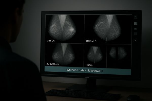
Digital breast tomosynthesis11 generates enormous datasets, and reviewing them efficiently is a major challenge. The traditional dual 5MP setup can feel cramped, forcing radiologists to constantly toggle windows or scroll through image stacks. Single-head 8MP and, particularly, 12MP displays12 are a powerful solution. Their vast screen real estate allows PACS hanging protocols to automatically arrange images in an optimized layout—for instance, displaying the CC and MLO tomosynthesis cine loops side-by-side, with the corresponding 2D synthetic images and priors arranged below or to the side. A 12MP monitor like the MD120C provides a seamless canvas for this, allowing the radiologist’s eyes to travel smoothly across all relevant information without needing to switch contexts or manipulate windows. This dramatically reduces wasted clicks and mouse mileage, accelerates reading times, and allows for more intuitive comparison between different views and examinations, directly boosting diagnostic throughput.
Color vs Grayscale in Mammography: When and Why
Believing mammography is purely grayscale overlooks modern needs. CAD markers, reports, and fusion imaging all require accurate color that a grayscale-only display cannot provide.
While grayscale is primary, high-quality color capability is increasingly important for viewing CAD overlays, reports, and fused multimodality images. A single color-capable display can handle every aspect of the modern mammography workflow.
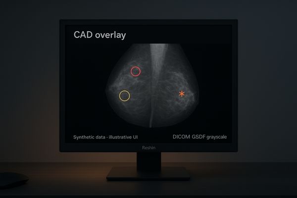
While the diagnostic information in a mammogram is found in its grayscale data, the modern workflow is not exclusively monochrome. Computer-aided detection (CAD) systems12 use brightly colored markers (circles, asterisks) to highlight suspicious areas. Patient reports, RIS/PACS interfaces, and even image processing tools often use color cues. Furthermore, as multimodality imaging grows, a radiologist may need to review a mammogram alongside a color-coded breast MRI or ultrasound image. A traditional grayscale-only display cannot render this information at all. For this reason, high-performance color diagnostic monitors13 are an excellent choice for mammography workstations. A display like the MD50C is engineered with a dual-LUT architecture: it delivers flawless, DICOM-calibrated grayscale for the primary image while simultaneously being able to render rich, accurate color for overlays and other applications. This provides the best of both worlds, creating a single, versatile workstation that can handle every task without compromise.
Ergonomics & Reader Fatigue: Layout, Eye Comfort, Speed
Hours spent staring at two separate screens with a bezel in between causes neck strain. Poor ergonomics and slow monitor response lead to fatigue and reduced reading accuracy over time.
Single-head 8MP/12MP layouts improve ergonomics by reducing head and eye movement. Anti-glare optics and flicker-free viewing further enhance eye comfort, while fast performance supports rapid, fatigue-free reading sessions.
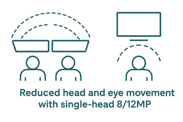
Radiologist burnout is a serious issue, and workstation ergonomics14 play a significant role. The traditional dual 5MP setup, with its central bezel, forces repetitive side-to-side head and neck movements, which can lead to chronic strain. Migrating to a single large-format 8MP or 12MP monitor eliminates the bezel and creates a more unified visual field, which studies have shown can reduce physical discomfort. Beyond the layout, the display’s optical properties are also key. Advanced anti-glare coatings reduce eye strain by minimizing distracting reflections without sacrificing image sharpness. Flicker-free backlighting technology provides a stable, comfortable viewing experience, which is essential during long reading sessions that involve scrolling through hundreds of DBT slices. By combining a more ergonomic layout with eye-friendly technology, these advanced displays help radiologists read more comfortably and efficiently, sustaining high performance throughout the day.
Integration with PACS & QA: Practical Deployment
A new monitor that doesn’t work with the existing PACS or quality assurance software is a failed investment. Seamless integration is essential for a smooth and compliant clinical operation.
Medical-grade displays are designed for seamless integration with PACS and QA software. Standardized connectivity and bundled QC tools ensure smooth deployment and simplify the process of maintaining regulatory compliance.
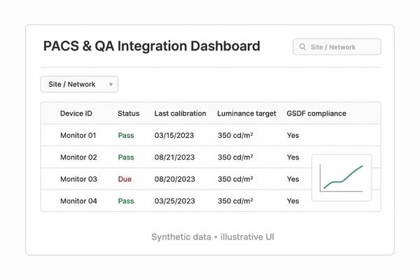
A mammography display is one component in a complex ecosystem that includes the imaging modality, the PACS (Picture Archiving and Communication System), and quality assurance (QA) software. For practical deployment, the monitor must integrate flawlessly. Medical-grade displays are designed with this in mind. They use standard DisplayPort connectivity, ensuring compatibility with modern graphics cards. Crucially, they are bundled with QA software15 that communicates directly with the monitor’s internal sensors and electronics. This software automates calibration checks, maintains a log of performance for regulatory requirements (like MQSA in the U.S.), and can often be managed remotely by a PACS administrator. This tight integration ensures that the monitor not only performs to spec out of the box but that its compliance can be easily verified and maintained over its entire lifecycle, making it a reliable and low-maintenance component of the clinical workflow.
Migration Path: From Dual 5MP to Single-Head 8/12MP
Upgrading an entire fleet of monitors to 12MP at once is often not feasible. A phased, logical migration strategy is needed to balance budget with clinical need.
A practical migration path involves replacing aging dual 5MP setups with 8MP single-head displays for general use. The highest-performance 12MP monitors can be reserved for high-volume DBT centers or lead radiologists.
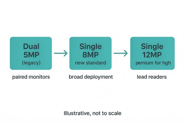
As older dual 5MP monitors reach their end-of-life, radiology departments have an opportunity to upgrade to more efficient technology. A complete, immediate switch to top-of-the-line 12MP display16s may not be financially practical for every institution. A sensible, phased migration strategy is often the best approach. The first step is to replace aging dual 5MP workstations with single 8MP displays17. A multi-modality display like the MD85CA is an excellent choice for this, as it provides a significant workflow improvement for both FFDM and DBT reading and can also be used for other radiological studies. This establishes a new, higher standard of care across the department. For centers with very high tomosynthesis workloads or for lead interpreting radiologists who demand the ultimate in efficiency, the investment in a 12MP display can be justified by its superior throughput. This tiered approach allows a department to modernize its fleet strategically, balancing budgetary constraints with the clear clinical benefits of next-generation display technology.
Conclusion
Reshin’s medical-grade mammography monitors provide the calibrated precision, stable performance, and workflow efficiency needed to meet the challenges of FFDM and DBT, supporting earlier and more reliable cancer detection.
👉 For expert guidance on selecting the right mammography display solutions, contact Martin at martin@reshinmonitors.com — we’re here to support your imaging needs.
-
Explore this link to understand how DBT enhances breast cancer detection and the technology behind it. ↩
-
Explore this link to understand the innovative technology behind 3D mammography and its benefits for breast cancer detection. ↩
-
Learn how CAD systems enhance the accuracy of mammograms by highlighting potential issues, improving early detection. ↩
-
Explore how 5MP monitors enhance image clarity and detail in mammography, crucial for accurate diagnoses. ↩
-
Learn about DBT’s advantages in mammography, including improved image quality and workflow efficiency for radiologists. ↩
-
Explore this link to understand how medical-grade displays enhance image quality and safety in mammography. ↩
-
Learn about brightness control technology to see how it ensures consistent performance and image clarity over time. ↩
-
Understanding GSDF is crucial for ensuring accurate mammographic interpretations and maintaining diagnostic quality. ↩
-
Understanding this technology is crucial for ensuring accurate mammography readings and preventing false positives. ↩
-
Exploring this topic will reveal how monitors maintain consistent performance over time, vital for medical imaging accuracy. ↩
-
Explore this link to understand how Digital breast tomosynthesis enhances breast cancer detection and improves diagnostic accuracy. ↩
-
Explore this link to understand how CAD systems enhance mammogram analysis and improve diagnostic accuracy. ↩ ↩
-
Discover the benefits of high-performance color diagnostic monitors for mammography workstations and their impact on patient care. ↩
-
Understanding workstation ergonomics can significantly enhance comfort and efficiency for radiologists, reducing burnout. ↩
-
Learn about the role of QA software in ensuring compliance and performance in medical imaging systems. ↩
-
Learn about the advantages of 12MP displays for high tomosynthesis workloads and efficiency in radiology. ↩
-
Explore how 8MP displays can enhance workflow and improve diagnostic accuracy in radiology departments. ↩


