Using the wrong display for a clinical task can have serious consequences. A slight inaccuracy can lead to a misdiagnosis. The right monitor is not just a screen; it is a precision instrument.
Medical displays are not one-size-fits-all. This article covers specific scenarios like diagnostic radiology, surgery, pathology, ICU monitoring, and telemedicine. We explain how each environment demands unique features like resolution, brightness, and color precision.
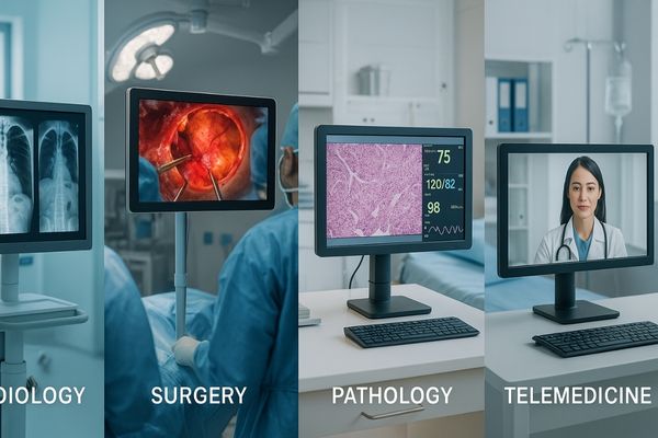
Different departments within a hospital have vastly different visual requirements. The way a radiologist examines a CT scan is not the same as how a surgeon views an endoscopic video. Each scenario presents unique challenges and demands a display technology1 engineered to meet them. Understanding these specific application needs is the first step toward equipping clinical teams2 with the tools they need to perform their best work. Let us look at some of the most common applications.
Diagnostic Radiology Requires High-Resolution and Grayscale Accuracy
A radiologist sees a subtle gray shadow on a scan. Is it a harmless artifact or a developing tumor? The wrong monitor makes this distinction nearly impossible, risking a critical misdiagnosis.
Diagnostic radiology depends on subtle grayscale detail. High resolution, stable brightness, and precise DICOM calibration are essential for making accurate interpretations of medical images like X-rays and CT scans.
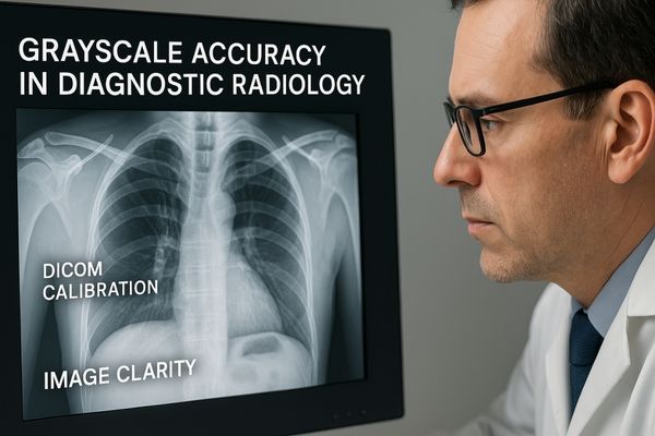
In diagnostic imaging, the ability to perceive minute variations in gray shades is paramount. The human eye can only distinguish a limited number of gray levels, so the display must render these details perfectly according to the DICOM Part 14 Grayscale Standard Display Function3. This standard ensures that an image looks the same on any compliant monitor. Consumer-grade monitors cannot meet this standard. They lack the necessary luminance stability and bit depth. As a result, they can either hide subtle pathologies or create visual artifacts that mimic them. A true diagnostic display must also have a higher resolution, often 3MP, 5MP, or even 12MP, to show the finest anatomical structures without forcing the user to pan and zoom constantly. This combination of resolution and grayscale precision, found in products like our MD52G – 5MP Grayscale Mammography Monitor4, gives radiologists the visual confidence they need to make life-saving decisions.
| Feature | Consumer Monitor | Medical Diagnostic Monitor |
|---|---|---|
| Grayscale Control | Basic brightness/contrast | DICOM Part 14 calibrated |
| Luminance | Unstable, drops over time | Stable, sensor-controlled |
| Resolution | Typically FHD or 4K | 2MP up to 12MP |
| Quality Assurance | None | Built-in QA, remote management |
Surgical Environments Demand Brightness and Real-Time Imaging
A surgeon performs a procedure under intense operating room lights. A standard monitor’s screen washes out completely. This glare and poor image quality can compromise surgical precision.
Operating rooms are brightly lit and fast-paced environments. Surgical displays must provide high brightness, effective anti-glare properties, and zero-latency imaging to support precise surgical actions.
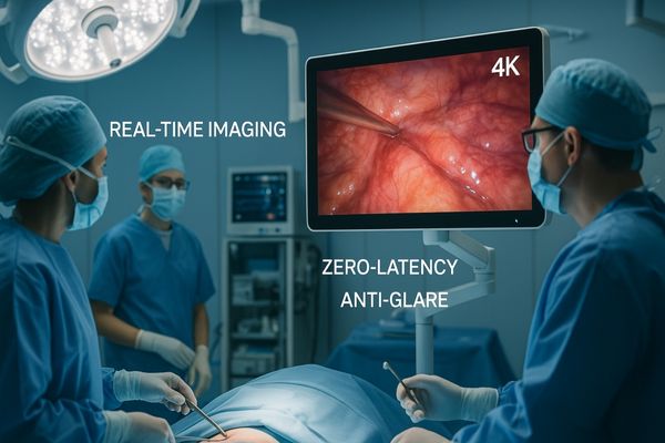
The operating room (OR) is one of the most demanding environments for any display technology. Ambient lighting from powerful surgical lamps can be extremely bright, easily overwhelming a conventional screen. Surgical displays must have a very high brightness output5, often exceeding 800 cd/m², to produce a clear, vibrant image that is not washed out. The screen surface also needs an advanced anti-glare and anti-reflection treatment. This ensures the surgical team can see the display clearly from any angle without distracting reflections. Furthermore, for minimally invasive procedures like endoscopy or laparoscopy, real-time imaging is non-negotiable. Any delay, or latency, between the surgeon’s actions and the image on the screen could have catastrophic consequences. Surgical monitors are engineered with advanced signal processing to ensure lag-free video6. The MS321PB – 32" 4K Surgical Monitor is built for this environment, combining high brightness, 4K resolution, and near-zero latency for optimal surgical performance.
Pathology and Dermatology Benefit from Color Precision
A pathologist examines a digital slide of a tissue biopsy. The colors of the chemical stains appear muted or shifted. This poor color fidelity could lead to an incorrect cancer grade.
Digital pathology and clinical dermatology rely on seeing true-to-life colors. Accurate and consistent color reproduction is essential for reliably identifying tissue stains and lesion characteristics for diagnosis.
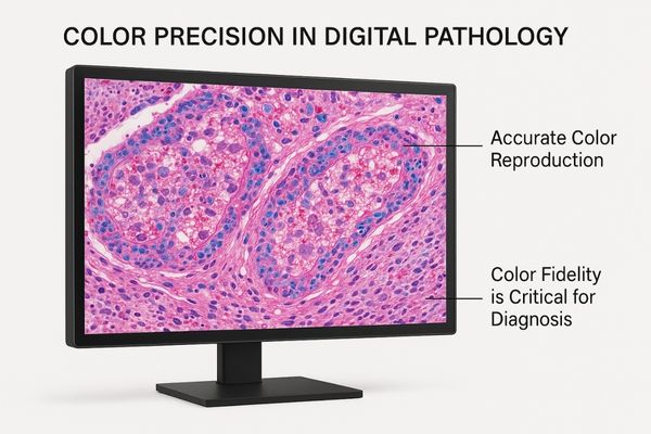
While radiology is a world of grayscale, disciplines like pathology and dermatology live in a world of color. Pathologists use specific chemical stains, like Hematoxylin and Eosin (H&E)7, which color cell nuclei blue and the cytoplasm pink. The precise hue, saturation, and intensity of these colors provide critical diagnostic information. A monitor that cannot accurately reproduce these colors is unsuitable for this work. Similarly, a dermatologist assessing a skin lesion on a screen needs to trust that the reds, browns, and blues they see are true to life. Medical color displays are designed to cover a wide color gamut, often encompassing 100% of the sRGB space or more. More importantly, they undergo a rigorous calibration process to ensure color accuracy and consistency over time. This guarantees that the colors on the screen faithfully represent the actual sample, supporting diagnostic confidence. Products like the MD50C – 5MP Color Mammography Monitor offer the color precision required for these demanding clinical applications.
| Field | Key Visual Information | Why Color Matters |
|---|---|---|
| Pathology | Stained tissue samples | Differentiates cell types, indicates disease state. |
| Dermatology | Skin lesions, rashes | Color and pattern are key to identifying conditions. |
| Endoscopy | Internal tissue | Distinguishes healthy from inflamed or abnormal tissue. |
Intensive Care Units Depend on Continuous Patient Monitoring
An alarm fails to display on a flickering ICU monitor. A patient’s vital signs are dropping unnoticed. This kind of equipment failure in a critical care setting can be fatal.
Intensive care units need to display vital signs 24/7. This demands medical monitors with exceptional reliability, durability, and strong resistance to electronic interference from nearby equipment.
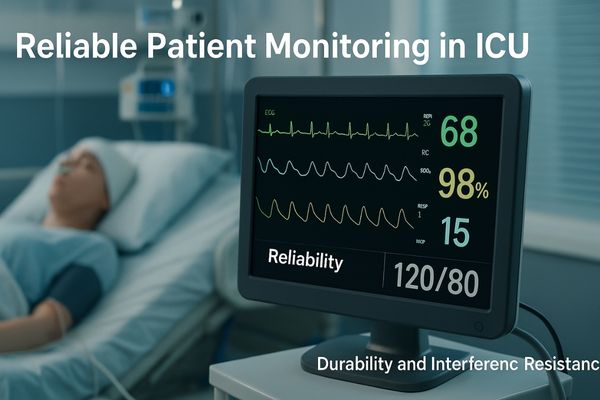
Monitors in the Intensive Care Unit (ICU) or at the patient’s bedside serve a different but equally critical function. Their primary job is to display a continuous stream of vital signs data, such as heart rate, blood pressure, and oxygen saturation. These are not diagnostic imaging displays, but their reliability is a matter of life and death. An ICU is a harsh electronic environment, filled with pumps, ventilators, and defibrillators that can generate electromagnetic interference (EMI). Patient monitoring displays must have robust internal shielding to prevent this EMI from disrupting their operation. They must also be built for 24/7 use with high-quality components and power supplies that will not fail unexpectedly. Furthermore, infection control is a top priority, so these displays need sealed, easy-to-clean housings that can withstand harsh chemical disinfectants. A versatile and reliable display like the MS192SA – 19" HD Endoscopic Monitor can be adapted for such patient monitoring cart applications due to its durable construction and medical-grade certifications.
Telemedicine Expands Need for Cross-Platform Consistency
A specialist reviews a patient’s case remotely. The MRI scan they see on their screen looks different from how it appeared to the local radiologist. This inconsistency creates diagnostic uncertainty.
The rapid growth of telemedicine makes image consistency across different platforms and devices essential. This is critical for enabling reliable remote consultations and effective collaboration between hospitals.

Telemedicine is changing how healthcare is delivered. It connects patients with specialists hundreds of miles away and allows for collaboration between clinicians at different institutions. For this to work safely and effectively, everyone must be looking at the same picture. If an image is viewed on a non-medical-grade monitor at one end and a calibrated diagnostic display at the other, the diagnostic information can be lost or distorted. This creates a weak link in the chain of care. True diagnostic confidence in teleradiology or telepathology requires that all viewing stations are calibrated to the same standard, typically DICOM8. This ensures perceptual consistency, meaning the image looks functionally identical regardless of the display location. This requires displays that not only support calibration but also have centralized quality assurance tools to verify their performance remotely. The MD45C – Dual-screen Diagnostic Monitor (Single Panel) is ideal for this, allowing clinicians to seamlessly compare current and prior images from different sources with guaranteed consistency.
Conclusion
The right medical display is a specialized instrument. Its design must match the demands of the specific clinical environment to ensure accuracy, reliability, and ultimately, patient safety.
📩 Need help choosing displays tailored to your clinical workflows? Contact Martin at martin@reshinmonitors.com for expert recommendations from Reshin.
-
Explore this link to discover cutting-edge display technologies that enhance medical imaging and improve diagnostic accuracy. ↩
-
Learn how tailored display solutions empower clinical teams to perform better, ensuring optimal patient care and outcomes. ↩
-
Understanding this standard is crucial for ensuring accurate diagnostic imaging and consistent image quality across monitors. ↩
-
Exploring the advantages of this monitor can enhance your knowledge of diagnostic imaging technology and its impact on patient care. ↩
-
Understanding high brightness output is crucial for ensuring clear visibility in demanding surgical environments. ↩
-
Exploring lag-free video technology can reveal its critical role in enhancing surgical precision and safety. ↩
-
Understanding H&E’s role in pathology can enhance your knowledge of diagnostic techniques and their importance. ↩
-
Understanding DICOM is crucial for ensuring image consistency in telemedicine, enhancing diagnostic accuracy. ↩