Pathologists spend hours examining slides, where every detail matters. The shift to digital workflows means that the monitor is now the microscope. An inadequate display can cause eye strain and diagnostic uncertainty.
The best displays for digital pathology review are medical-grade monitors with high resolution (at least 4MP, ideally 5MP to 8MP), exceptional color accuracy (10-bit depth, BT.709/sRGB coverage), and a large screen size (32 inches or greater) for efficient workflow.
A diagnostic monitor for digital pathology should provide high resolution (4MP–8MP+), calibrated wide-gamut color, uniform brightness, and a large screen area. These features allow pathologists to interpret WSI slides with clarity, maintain diagnostic confidence, and reduce eye strain during long review sessions.
As an engineer who has deployed diagnostic displays for WSI and high-volume pathology labs, I’ve repeatedly seen how the right monitor directly affects diagnostic speed, fatigue, and accuracy. Below, I summarize the core requirements that matter most in digital pathology display selection.
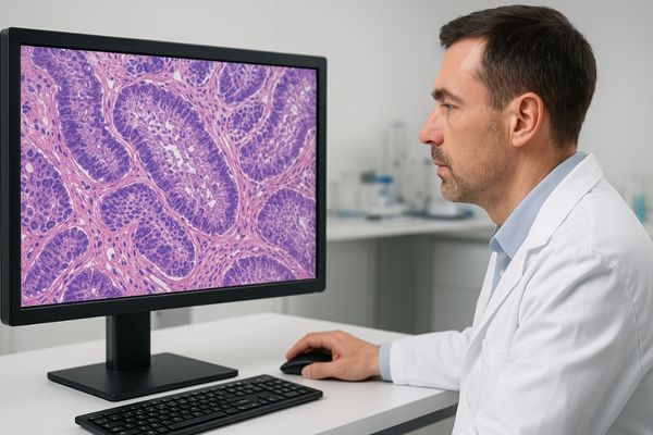
The transition from traditional glass slides to digital whole slide imaging (WSI)1 has revolutionized the field of pathology. This technology allows for easier collaboration, archival, and analysis. However, it also places new and intense demands on the displays used for review. A consumer-grade monitor is simply not built for the rigor and precision required for making a diagnosis from a digital image. The right display is a critical instrument that directly impacts diagnostic confidence and efficiency.
In my digital pathology display integration work, I often start by evaluating the core visual requirements of the workflow. We will discuss resolution2, color accuracy3, uniformity, and ergonomics so you can understand exactly what to look for in a display capable of meeting these challenges.
What visual demands are unique to digital pathology review?
Digital pathology is more than just viewing a static image. It is an active process of navigation and analysis. A standard office monitor can cause eye fatigue and frustration during this dynamic work.
Digital pathology uniquely demands exceptional clarity for constant zooming and panning across massive whole slide images. It requires precise color fidelity to interpret stains correctly, fast pixel response for motion stability, and ergonomic features like uniform brightness, anti-glare surfaces, and flicker-free operation.
In my deployments for pathology workstations, I’ve seen how the unique nature of navigating gigabyte-scale WSI datasets introduces several engineering-level requirements:
- Fast pixel response time is essential to prevent motion blur when panning across dense cellular regions.
- Brightness and color uniformity must meet medical-grade tolerance to prevent misinterpretation caused by hot spots or dim corners.
- Ergonomic comfort—including flicker-free backlights, low-blue-light modes, and strong anti-glare performance—is critical for long review sessions.
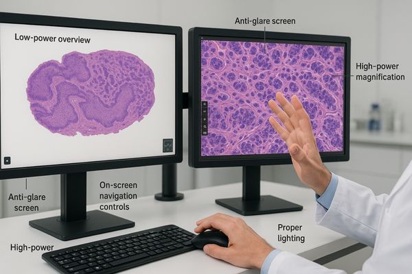
The MD45C – Dual-screen Diagnostic Monitor (Single Panel) supports this workflow by allowing a pathologist to view a WSI alongside patient data or comparative slides on a seamless 4MP surface.
How much resolution is required for accurate pathology diagnosis?
In my field evaluations, resolution is often the first limiting factor that pathologists notice. Using a low-resolution screen is like looking through an out-of-focus microscope—it hides crucial cellular detail.
A minimum resolution of 4MP is recommended for digital pathology. For primary diagnosis or complex cases, 5MP to 8MP displays are superior because they show more detail at once, reducing excessive zooming and boosting diagnostic speed and confidence.
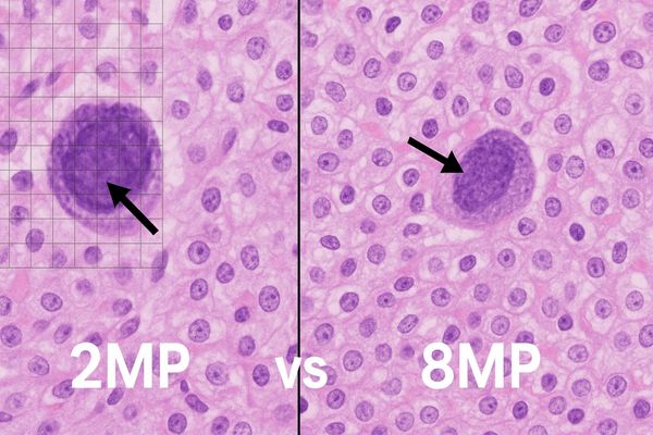
Resolution directly determines how much of the slide a pathologist can see at once without sacrificing detail. A higher resolution4 allows users to maintain context while still identifying fine cellular structures—a major workflow advantage.
For example, on an 8MP display, a pathologist can assess tissue architecture while simultaneously examining nuclear morphology. This reduces constant zooming and panning, lowering cognitive load.
While 4MP is a functional entry point, stepping up to 5MP or 8MP is a direct investment in diagnostic quality5. It reduces the risk of missing small foci of disease less apparent at lower resolutions.
Our MD51CHY – 34" 5MP Diagnostic Monitor provides exactly this advantage, making it well-suited for detailed pathology review.
Recommended Resolution for Digital Pathology
| Resolution | Pixel Dimensions | Best Use Case |
|---|---|---|
| 2MP (FHD) | 1920 x 1080 | Not for diagnosis; suitable for lab information systems. |
| 4MP (QHD) | 2560 x 1440 | Minimum recommended for routine review. |
| 5MP | 2560 x 2048 | Primary diagnosis and detailed evaluation. |
| 8MP (4K) | 3840 x 2160 | Ideal for complex cases & high-volume labs. |
| 12MP+ | 4200 x 2800+ | Top choice for multi-slide comparison, research, or advanced WSI. |
Do color accuracy and wide gamut significantly impact pathology work?
In my experience calibrating lab workstations, color accuracy is one of the most underestimated factors. Stains must look exactly as intended for confident interpretation.
Yes, color accuracy is critical. Histological stains contain diagnostically meaningful color variations. Wide-gamut and 10-bit color support ensure subtle hues—especially in stains like H&E—are reproduced accurately and consistently.
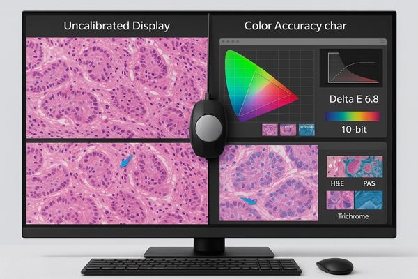
Pathology relies heavily on interpreting stain color. Stains such as Hematoxylin and Eosin (H&E)6 require precise hue and saturation reproduction.
From an engineering perspective, there are three non-negotiable requirements:
1. Wide color gamut (100% sRGB / BT.709 or higher)
Consumer monitors often miss pastel tones found in cytoplasm, or deep blues in nuclei.
2. True 10-bit color depth + 10-bit LUT
A medical display shows 1.07 billion colors, revealing gradients an 8-bit display cannot.
3. Hardware calibration
Displays drift over time. Hardware LUT calibration ensures consistent long-term performance.
The MD50C – 5MP Color Diagnostic Monitor delivers pathology-appropriate color uniformity, calibration capability, and contrast precision.
In pathology labs I’ve supported, navigation fatigue is one of the most common complaints. Constant panning slows the workflow and contributes to eye strain—but the right screen can correct this.
A large screen (32"+) paired with high pixel density gives pathologists more context at once, reducing navigation effort. High PPI keeps details sharp even at lower zoom levels.
![]()
Screen size and pixel density7 work together:
- A larger canvas reveals more tissue architecture.
- A higher PPI produces microscope-like clarity.
- High resolution enables efficient overview + detail review.
This combination transforms WSI navigation from tedious to efficient.
Our 8MP Diagnostic Display provides a large-format 4K surface ideal for pathology.
Impact of Screen Size on Viewable Area (equal magnification)
| Screen Size | Relative Area | Workflow Impact |
|---|---|---|
| 24" | 1.0x | Heavy panning. |
| 27" | ~1.3x | Better context, less panning. |
| 32" | ~1.8x | Excellent overview; major workflow boost. |
Which Reshin displays are designed for digital pathology use?
Over years of supporting digital pathology transitions, I’ve seen that choosing the right hardware is essential—not only for image clarity but for long-term workflow stability.
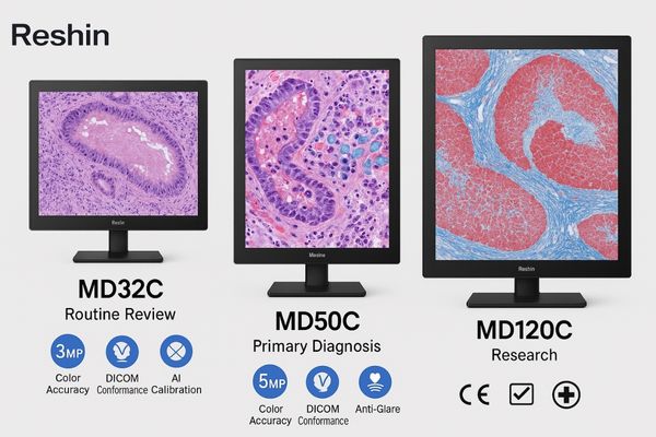
Reshin offers a dedicated line of diagnostic displays for digital pathology—including MD50C (5MP Color), MD51CHY (5MP), and the flagship MD120C (12MP). These displays provide calibrated color, high resolution, uniformity compensation, and ergonomic features built for long review sessions.
Based on our deployment experience across diagnostic labs:
- MD50C (5MP Color): best for color-dependent workflows (H&E, IHC) requiring wide-gamut fidelity.
- MD51CHY (5MP): balanced choice for detailed WSI review in mid- to high-volume labs.
- MD120C (12MP): optimal for large pathology centers requiring simultaneous slide comparison or multi-panel workloads.
All Reshin displays support:
- DICOM Part 14 conformance8
- Factory calibration + hardware LUT recalibration
- Anti-glare coatings, flicker-free backlights, low-blue-light ergonomics
- Strong brightness uniformity and compensation
For the most demanding pathology environments, the MD120C brings 12MP resolution + AI calibration9, enabling full-slide viewing with unparalleled precision.
FAQ
1. Can I use a consumer monitor for digital pathology?
Not for diagnosis. Consumer displays lack calibrated color, uniformity control, 10-bit support, and brightness stability—leading to missed details and fatigue.
2. Is 4K enough for primary pathology review?
4K (8MP) is excellent, but 5MP color displays remain the most balanced choice for stain interpretation and cellular detail.
3. Why is 10-bit color essential?
10-bit LUT + wide gamut ensures accurate reproduction of stain gradients that may indicate disease severity.
4. Do pathology displays need DICOM Part 14 compliance?
Yes. Even though pathology is color-driven, DICOM Part 14 ensures consistent brightness and gamma across multi-monitor environments, supporting reliable long-term WSI review.
Conclusion
Choosing a medical-grade monitor with high resolution, precise color, and a large screen is not optional for digital pathology. It is essential for diagnostic accuracy, workflow efficiency, and pathologist well-being.
In our field projects, we consistently help pathology teams evaluate display performance based on WSI resolution, stain fidelity, and workflow requirements.
To upgrade your digital pathology workflow with purpose-built diagnostic displays, you can contact Reshin’s engineering team directly — we are ready to support your project needs.
📧 Email: info@reshinmonitors.com
🌐 Website: https://reshinmonitors.com
-
Overview of how WSI technology enhances collaboration and analysis. ↩
-
Why resolution matters for pathology detail and workflow. ↩
-
Explanation of color accuracy and its diagnostic impact. ↩
-
How higher resolution improves diagnostic clarity. ↩
-
Study on resolution’s role in diagnostic confidence. ↩
-
Insight into H&E staining and its diagnostic relevance. ↩
-
How PPI affects pathology clarity. ↩
-
Meaning of DICOM Part 14 conformance in diagnostics. ↩
-
Benefits of AI-based calibration in diagnostic displays. ↩