Inaccurate color on a medical display can lead to misdiagnosis. This compromises patient safety and makes clinical decisions difficult. Selecting a display with certified color accuracy is the solution.
This guide explains how to evaluate color accuracy in medical displays. We will cover key specifications, calibration, clinical needs, and how to balance cost with performance for the best clinical value.
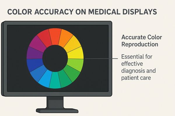
Choosing the right medical display is a critical decision. Color performance is not a luxury; it is a fundamental requirement for safe and effective patient care. Let us explore the key factors that ensure you are selecting a display with the color accuracy your institution needs.
Color Accuracy Impacts Diagnostic Confidence and Outcomes
Subtle color variations in a medical image can obscure vital information. This uncertainty can lead to missed pathological details or incorrect tissue identification during surgery. High color accuracy is essential.
In medical imaging, even slight color deviations can lead to the misidentification of lesions or tissues. High color accuracy is therefore a non-negotiable requirement for confident clinical diagnosis and safe procedures.
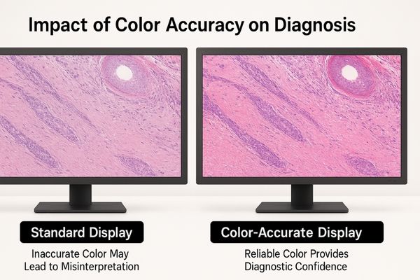
The human eye is incredibly sensitive to color, and clinicians rely on this ability to interpret complex visual information. In fields like digital pathology, the precise shades of hematoxylin and eosin (H&E) stains1 reveal cellular structures and abnormalities. If a display renders these colors incorrectly, a pathologist might misinterpret the tissue. Similarly, during an endoscopic procedure, the surgeon must differentiate between healthy and unhealthy tissue based on subtle variations in redness and texture. Inaccurate color can mask these differences, potentially affecting surgical margins or decisions. This is why a display’s ability to reproduce colors exactly as they are captured is directly linked to diagnostic confidence and patient outcomes. The MS321PB – 32" 4K Surgical Monitor is designed to deliver this level of fidelity, ensuring surgeons see tissue in true-to-life color, which is critical for procedural precision.
Key Specifications Define Display Color Performance
Display specification sheets are often filled with technical jargon. Choosing a monitor based on the wrong information can lead to poor color performance in a clinical setting. Focusing on a few key metrics simplifies the decision.
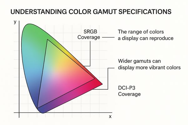
When buying a medical display, focus on key parameters. These include color gamut coverage like sRGB or DCI-P3, grayscale performance for DICOM compliance, and Delta E values for color deviation.
To make an informed choice, you must understand three core specifications that define a display’s color capabilities. These metrics provide objective data about how well a monitor can reproduce color and grayscale tones.
Critical Color Specifications
- Color Gamut2: This refers to the range of colors a display can produce. It is often expressed as a percentage of a standard color space, such as sRGB or DCI-P3. A wider gamut means the display can show more vibrant and saturated colors, which is important for applications like endoscopy.
- Grayscale Linearity (DICOM)3: For radiology, the ability to show distinct shades of gray is critical. A display must comply with the DICOM Part 14 standard to ensure that grayscale images are consistent and diagnostically accurate.
- Delta E (ΔE): This value measures the difference between a source color and the color a display reproduces. A ΔE value of less than 2 is generally considered undetectable by the human eye, indicating high color accuracy.
The MD50C – 5MP Color Mammography Monitor excels in these areas, offering a wide color gamut and low ΔE for precise color pathology and mammography fusion imaging.
| Specification | What It Measures | Why It Matters |
|---|---|---|
| Color Gamut (% sRGB/DCI-P3) | The range of colors the display can show. | A wider gamut provides more vivid, true-to-life colors for endoscopy and pathology. |
| DICOM Part 14 Conformance | Consistency in displaying shades of gray. | Essential for accurate diagnosis from X-rays, CT scans, and MRIs. |
| Delta E (ΔE) | The difference between input color and displayed color. | A low value (ΔE < 2) signifies high color fidelity, crucial for all clinical tasks. |
Calibration Capabilities Ensure Long-Term Consistency
All displays experience color and brightness drift over time. This gradual degradation can happen unnoticed, silently compromising diagnostic quality and consistency. Onboard calibration features solve this problem effectively.
Displays with hardware or software calibration capabilities maintain stable color performance over their lifespan. This ensures consistent diagnostic quality and reduces the need for frequent manual service and maintenance.
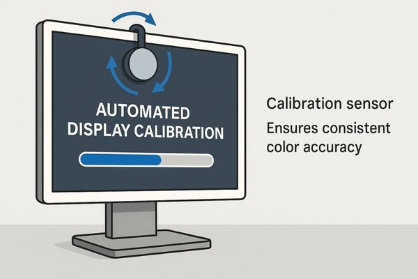
A display may be perfectly color-accurate when it leaves the factory, but its performance will change with use. This is due to the natural aging of the backlight and other components. Calibration is the process of measuring the display’s output and adjusting it back to a known standard. This step is essential for maintaining diagnostic confidence over the long term. Medical displays offer different types of calibration4. Software calibration uses an external sensor and adjusts the computer’s video card. Hardware calibration uses a built-in sensor to directly adjust the display’s internal lookup table (LUT), providing more precise and stable results. The MD26GA – 2MP Diagnostic Monitor features an integrated front sensor for automated DICOM compliance5 checks and calibration, ensuring it remains diagnostically accurate with minimal human intervention. This automated process saves time for IT staff and gives clinicians peace of mind.
Clinical Scenarios Determine Color Accuracy Requirements
A single type of medical display cannot meet the needs of every department. Using a display that is not optimized for its specific clinical task can hinder workflow and even compromise care. Matching the display to the application is key.
Operating rooms, radiology reading rooms, and pathology labs each have unique color accuracy needs. The selection of a display must be tailored to the specific visual requirements of each clinical scenario.
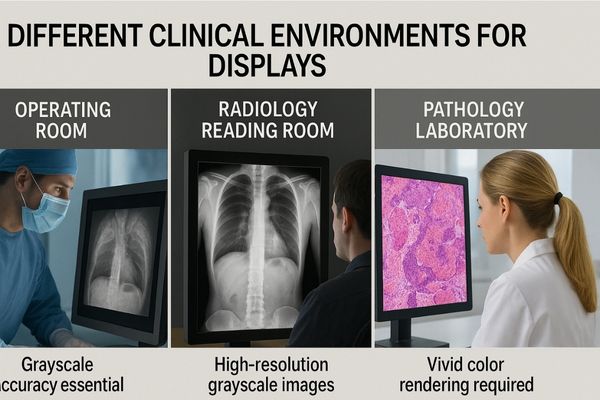
The "best" display is always the one that is best for the job. Different medical disciplines have different visual priorities, and a smart procurement strategy accounts for these variations.
Matching Displays to Departments
- Radiology: This field demands exceptional grayscale performance. Radiologists need to perceive subtle differences in tissue density on X-rays, CTs, and MRIs. Therefore, DICOM Part 14 conformance6 is the most important feature.
- Pathology: Pathologists rely on true-to-life color to interpret stained tissue samples. A display with a wide color gamut (covering 100% sRGB) and a low Delta E is essential for distinguishing cellular details accurately.
- Surgery and Endoscopy: In the operating room, surgeons need to see vibrant, accurate color in real-time to differentiate anatomical structures and identify blood flow. A 4K display with a wide color gamut7, high brightness, and excellent video processing is required.
The MD33G – 3MP Diagnostic Monitor is an excellent example of a display built for a specific need. Its high-resolution grayscale panel is optimized for chest X-rays and other radiological studies where DICOM compliance is paramount.
| Clinical Area | Primary Visual Task | Key Display Requirement |
|---|---|---|
| Radiology | Differentiating tissue densities | DICOM Part 14 Grayscale Conformance |
| Pathology | Interpreting stained tissue slides | High Color Fidelity (Low ΔE, sRGB Gamut) |
| Surgery | Identifying live tissue and anatomy | 4K Resolution, Wide Color Gamut, Low Latency |
Balancing Cost and Performance in Purchase Decisions
High-performance medical displays often represent a significant investment. Strict budget constraints can tempt procurement teams to make compromises that might affect clinical quality. A smart evaluation focuses on total value, not just the initial purchase price.
Balancing budget constraints with the need for diagnostic-grade color accuracy is essential. This ensures the hospital achieves the best long-term value and maintains high standards of clinical performance.
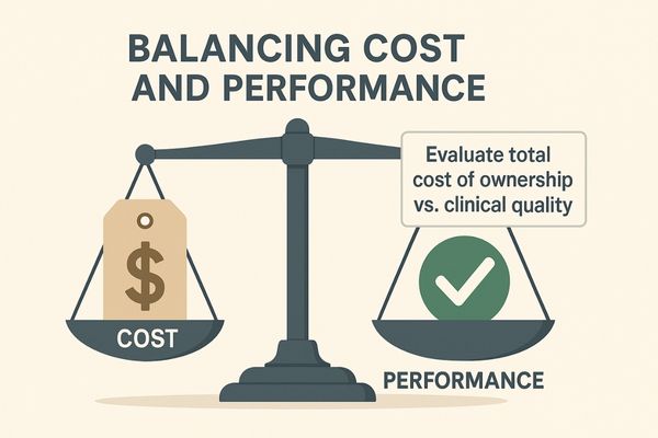
The upfront cost of a medical display is only one part of the equation. A cheaper, consumer-grade monitor may seem like a good deal, but it can lead to higher costs over its lifetime. This is where the concept of Total Cost of Ownership8 (TCO) becomes important. TCO includes not only the purchase price but also factors like lifespan, warranty, service costs, and the need for frequent replacement. A medical-grade display9 is built with higher-quality components designed for longevity and reliability in a demanding 24/7 environment. It also comes with the necessary certifications for clinical use, which prevents compliance issues. Investing in a durable, reliable display with a strong warranty, like the MS247SA – 24" FHD Endoscopic Monitor, can result in lower TCO and greater peace of mind. It provides the necessary performance for many procedures while offering excellent long-term value.
Conclusion
Color accuracy is a pillar of diagnostic confidence and procedural safety. Understanding key specifications, the role of calibration, and specific clinical needs allows for an informed and strategic purchasing decision.
📧 Need guidance on selecting displays with precise color performance for your clinical setting? Contact Martin at martin@reshinmonitors.com to learn more about Reshin’s solutions.
-
Understanding H&E stains is crucial for interpreting tissue samples accurately, enhancing diagnostic skills. ↩
-
Understanding Color Gamut is essential for evaluating display quality, especially in fields requiring color precision. ↩
-
Exploring Grayscale Linearity helps ensure accurate medical imaging, crucial for effective diagnosis in radiology. ↩
-
Understanding calibration is crucial for maintaining display accuracy, especially in medical settings where precision is vital. ↩
-
Exploring DICOM compliance will enhance your knowledge of standards that ensure medical images are accurate and reliable. ↩
-
Understanding DICOM Part 14 conformance is crucial for radiologists to ensure accurate image interpretation and compliance. ↩
-
Exploring the concept of wide color gamut can enhance your knowledge of display technology and its impact on accurate tissue analysis. ↩
-
Understanding TCO helps in making informed decisions about medical equipment investments, ensuring long-term savings. ↩
-
Exploring the advantages of medical-grade displays can enhance your knowledge on reliability and compliance in clinical settings. ↩


